
One would think that being extraordinarily flexible would be advantageous to a dancer. After all, many forms of dance are characterized by extreme ranges of motion, particularly in the spine and lower extremities. But acrobatic flexibility may in fact be a sign of a system-wide disorder affecting the body’s connective tissues that can cause a host of symptoms ranging from chronic joint pain and other musculoskeletal complaints to, in more extreme cases, cardiovascular symptoms, digestive distress, and autonomic dysfunction.
In this article, I will discuss benign joint hypermobility syndrome (BJHS) – its etiology, symptoms, and diagnosis – and differentiate it from other forms of hypermobility. I will describe how BJHS presents in dancers and present general guidelines for manual and movement interventions appropriate in the context of a Rolfing® Structural Integration (SI) session.
Categories of Joint Hypermobility
Joint hypermobilities exist along a spectrum. People can be mildly affected and have few to no symptoms, or they can be severely affected and require braces or surgery to stabilize their joints. Dancers, particularly professional dancers, tend to fall on the more functional end of the spectrum, because the extraordinary athletic demands of professional dance generally prohibit individuals with pronounced symptoms from being successful onstage in the long term.
At the most functional end of the spectrum of joint hypermobilities is generalized joint hypermobility (GJH), which is characterized by an increased range of motion (ROM) relative to the general population in one or more joints. It is present in an estimated 5%-15% of the population and is more common among children, adolescents, and women, and among certain ethnic groups, particularly people of Asian and African descent. Individuals with GJH are frequently asymptomatic or have only mild symptoms. In fact, GJH may even be advantageous in dance, gymnastics, music, and sports.BJHS, the focus of this article is distinct from GJH. It is believed to be an inherited connective-tissue disorder, and it is characterized by increased joint ROM accompanied by chronic joint pain and other signs and symptoms related to a defect in the structure, production, or processing of collagen or the proteins that interact with collagen. As Rolfers, we are well aware that collagen, as the main structural protein of connective tissue, provides structure and support to body tissues. In people with BJHS, abnormal collagen renders these tissues more elastic. As a result, these individuals are at increased risk of joint instability due to ligament laxity, and they frequently present with strains, sprains, tears, and dislocations or subluxations. Other connective tissues can be hyperextensible as well, including tissues that support the skin and viscera. This may result in overly thin and stretchy skin, easy bruising, uterine or rectal prolapse, and hernias.
At the far end of the spectrum of joint hypermobilities are complex systemic disorders such as Ehlers-Danlos Syndrome (EDS)1 and Marfan Syndrome (MFS). This category of hypermobilities is rare and disabling and affects multiple body systems. Ligament laxity can be severe, and some individuals may require bracing and surgery. Like people with BJHS, these individuals may also have skin fragility and organ prolapse. Additional symptoms are related to increased tissue elasticity in the blood vessels and gut: cardiovascular manifestations may include low blood pressure, orthostatic intolerance, and mitral valve prolapse; digestive symptoms may include acid reflux, delayed gastric emptying, and irritable bowel syndrome. Autonomic nervous system dysfunction is common as well, because the body compensates for chronically low blood pressure and the associated fatigue by producing excess adrenalin.
So the spectrum of joint hypermobilities is quite vast and includes, on the one end, enhanced flexibility that may actually be of benefit in dance and athletics and, on the other end, systemic and disabling forms of hypermobility such as EDS (see Figure 1).
Hypermobile dancers generally fall into the first two categories; EDS and other severe forms of hypermobility are so limiting that they would prohibit a career in dance.
BJHS Among Dancers
An estimated 10% of the general population has BJHS, the more moderate form of joint hypermobility represented by the middle tier in Figure 1, but the prevalence among dancers is estimated to be as high as 44%- 58%.
In a study of dancers with the Royal Ballet School and Company in London (McCormack et al 2004), common musculoskeletal complaints among those identified as having BJHS included neck and lower back pain, ankle sprain, ligament injuries, fractures, and dislocations. Importantly, this study determined that not only were dancers with BJHS at higher risk of pain and injury than their non- BJHS counterparts, but also they were significantly more likely to take time off from dancing because of injury. One-half of the female dancers in the study who had BJHS reported having one or more tendon injury over a five-year period, and 61% of them stopped dancing for more than six weeks as a result. Among male dancers, 42% of those with BJHS reported tendon injury, while only 8% of those without BJHS suffered the same injury. Of those male dancers with BJHS with a tendon injury, a staggering 83% had to take six or more weeks off of dancing.
In a five-year follow-up to this study, the authors suggested that the tendons of dancers with BJHS may actually be not only weaker structurally due to collagen deficiency but also slower to strengthen in response to training and slower to heal as well. “The dancer with BJHS is both more vulnerable to the effects of injury and . . . healing is likely to be more prolonged and may be incomplete,” the authors conclude (McCormack et al 2009, 1613). A 2011 review of the research reached the same conclusion: “The modest amount of research on injury rates in hypermobile dancers confirms that they have substantially more tendon injuries and longer healing times than normal dancers (Day et al 2011, 488).”
Given that soft-tissue injury can cause dancers significant professional disability, it behooves those of us who work with dancers to be able to recognize BJHS and intervene early and appropriately.
Screening for BJHS
Professional companies screen new company members for joint hypermobility to determine their degree of joint laxity and ensure that they receive appropriate support behind the scenes. While most companies still use the Beighton score as a diagnostic tool, the Brighton criteria are now widely recognized as the most appropriate tool for identifying BJHS. Both of these tests are easy to perform and can be easily integrated into a Rolfing session.
Beighton Score
The Beighton score is an indicator of the most common form of hypermobility – GJH. A high Beighton score by itself does not mean that an individual has BJHS; other signs and symptoms must also be present.
The Beighton score is calculated as follows:
Brighton Criteria
The Brighton criteria were developed in 1998 and today are considered the diagnostic criteria for BJHS. Brighton uses the Beighton score along with other signs and symptoms to confirm diagnosis of BJHS.2 Major and minor Brighton criteria are as follows:
Major Criteria
Minor Criteria
A diagnosis of BJHS, which can only be given by a qualified medical professional, requires the presence of two major criteria, one major and two minor criteria, four minor criteria, or two minor criteria and an affected first-degree relative.
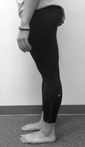
Figure 2: Hyperextended knees indicate GJH, according to the Beighton criteria. In combination with other signs and
symptoms, they may indicate BJHS.
Interventions
Broadly speaking, interventions for BJHS should seek to restore dynamic support to the hypermobile structures and, if appropriate, mobilize structures that are hypomobile relative to the hypermobility.
As Rolfers, we are comfortable recognizing joint hypomobilities and associated soft-tissue restrictions. Even in the BJHS dancer, there is likely to be stiffness elsewhere in the system as the body attempts to compensate for the hypermobility. There may be excess ROM in one lumbar segment relative its neighbor, for instance, or pronounced flexibility in the lumbars with corresponding stiffness in the thoracic spine. Where the hyper- and hypomobilities appear will vary from dancer to dancer and according to the style of dance – whether ballet, hip hop, Indian dance, etc. – and the choreographic demand of the pieces in active repertory. In Swan Lake, for instance, the principal ballerina performs many dozens of dramatic backbends that can destabilize the lumbar spine, and dancers in the Broadway musical The Lion King wear elaborate headpieces that can challenge cervical stabilization.
When there is limited range at one or more joints relative to neighboring joints, the hypermobile structures get exploited. Therefore, employing fascial release techniques to help mobilize the restricted structures is a critical. However, it is equally important to address the excessive mobility in the BJHS dancer. This is done by awakening the local muscles that stabilize the joint, thereby restoring dynamic support.
Movement Interventions
A healthy joint has both static and dynamic support. Static support is provided by appropriate stiffness in the surrounding ligamentous structures and also by the structure of the joint itself. Many dancers are born with joint structures that allow for excessive mobility – for instance, a retroverted femoral head, laterally oriented acetabulum, and long femoral neck allow for exaggerated hip external rotation. When naturally mobile joint structures are combined with ligament laxity, as is the case in BJHS, the job of supporting the joint is left largely to the musculature. Dancers with BJHS, therefore, unlike their non-BJHS counterparts, rely heavily on dynamic support from local muscles (sometimes called ‘local stabilizers’ or ‘postural muscles’) to maintain joint integrity. Given dancers’ grueling schedules, which can include as many as eight hours a day or more in the studio or onstage, it is easy to understand how local stabilizers can fatigue and how pain and injury can result.
Local muscles have a small movement arm and generate less torque and are therefore at a biomechanic advantage, relative to global muscles, for stabilizing joint structures. Familiar local stabilizers include multifidus, transversus abdominis, diaphragm, pelvic floor, rotator cuff muscles, and deep cervical flexors, to name a few. But technically any muscle is capable of providing stabilization in some capacity, depending on the demand. Ultimately, it is the nervous system working in concert with the muscular and articular systems that controls joint stabilization and movement.
Generally, local stabilization, when inhibited, is best rehabilitated through movement interventions. Such interventions should challenge the stability of the hypermobile structures with exercises that use little or no resistance and a high number of repetitions at a slow speed with a small range of motion (Figure 3). Using too much resistance results in the global muscles overriding the smaller muscles of local support. And since local stabilizers, composed largely of slow-twitch (type II) muscle fibers, are designed to be lightly active over a long period of time, low-intensity exercises that train endurance rather than speed are most effective.
Keys to Awakening Local Stabilizers
? Low load
? Small range of motion
? Slow speed
? High repetitions
? Proprioceptive challengeFigure 3: Movement interventions that seek to restore dynamicsupport should apply
Proprioceptive training is also critical to restoring local stabilization, since hypermobile joints have diminished proprioceptive acuity. Exercises performed on an unstable surface – such as a Bosu®, Tuning Board™, wobble board, or foam roller – enrich the client’s sensory experience and thereby help awaken joint proprioception. Closed kinetic chain exercises (in which the distal extremity meets resistance) also facilitate proprioceptors and stimulate co-contraction of the muscles around the joint, enhancing stability. Other benefits to closed-chain exercises are that they put less strain on the joints and ligaments than open-chain exercises (in which the distal extremity moves freely), and they better simulate functional demand.
There are an infinite number of ways to train local stabilizers in a Rolfing session, and the intervention will vary considerably from one dancer to the next according to her unique strengths and vulnerabilities.
Manual Interventions
It is often helpful to begin the work of rehabilitating local stabilization by intervening manually while the dancer’s extremities are in contact with a solid surface (closed chain). This could take a variety of forms – for instance, hands or feet on the wall, feet resting flat on the treatment table (hook-lying position), or work with the dancer in standing or quadruped (hands and knees) position. Using props similarly enhances feedback to the joints – for instance, having the dancer’s hand in contact with a ball when intervening at the shoulder girdle, putting a ball between her knees with legs in hook-lying position to enliven her medial line while working with the arches of the feet, and so on. Rolfers who have pursued Rolf Movement® Integration training will be familiar with many of these techniques and will be comfortable improvising. The underlying concept is to feed information to the dancer’s nervous system by enriching her sensory environment.
Exercise Interventions
Rolfing sessions with BJHS dancers should always include an exercise intervention, however brief, at the end of the session to awaken the deep stabilizers and integrate neuromuscular control. A simple intervention for the pelvis and lower extremities is one that most of us were exposed to in our basic Rolfing training: standing on Darrell Sanchez’s wonderful tool, the Tuning Board. Invite the dancer to balance in parallel or ‘turnout’ (hip external rotation), depending on the goal of your treatment session, on the Tuning Board or another unstable surface. Give her time to settle into the new sensation. Add challenge to the exercise, as appropriate, by asking her to slowly shift her balance to one leg or the other, to close her eyes with one or both hands lightly touching the wall for feedback, and so on. Be creative, keep movement slow and small, and draw upon the dancer’s movement vocabulary so that the lesson translates to her functional movement environment in the studio and onstage.
For Rolfers trained in Rolf Movement Integration or another movement practice like Pilates, Alexander Technique™, or the Feldenkrais Method® of somatic education, exercises can be more tailored in order to target hypermobile structures more specifically. Figure 4 shows an exercise intervention for a dancer with poor lumbopelvic control into hip flexion. The dancer lies supine on a foam roller and finds her balance with feet contacting the floor (A). Arms can be resting on the floor for support, resting on the ASISs to provide feedback about pelvic orientation (anterior tilt, posterior tilt, neutral), or moving freely for added challenge. Next, maintaining the natural lumbar curve and keeping ASISs level, the dancer marches one leg off the floor (B). The beauty of using the foam roller is that feedback is immediate: Without the support and coordination of the deep lumbopelvic stabilizers, the foam roller will roll and sway. The dancer will make micromovements below the level of conscious awareness to adjust in response to the feedback. The movement at the hip joint can vary according to the dancer’s ability to maintain neutral hips and spine. If stability is extremely compromised, she can begin by just lifting one heel away from the floor. For added challenge, she can gradually make the movement larger, extend the knee, incorporate hip external rotation, gesture the arms, and so on. Other variations include tying a TheraBand™ around the thighs to cue isometric activity of hip abductors, placing a ball between the knees to recruit adductors, and reaching arms overhead to involve upper abdominals, to name a few. Invite the dancer to perform the exercise right and left, comparing the more affected side with the more stable side, and encourage her to work slowly and with focused attention.
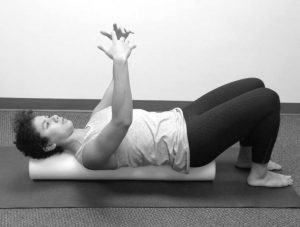
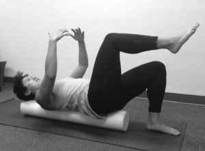
Figure 4: Dancer Nicole Turchi performs exercises on an unstable surface to activate lumbopelvic stabilizers.
Conclusion
In summary, joint hypermobility is common among dancers and may be a sign of a collagen defect. BJHS is characterized most prominently by pain, injury, and prolonged healing, and therefore can seriously impact a dancer’s career. BJHS is diagnosed by a qualified medical practitioner using the Brighton criteria, and Rolfers’ manual and movement interventions have the potential to alleviate symptoms by enhancing joint proprioception and training local stabilization.
Special thanks to Jena Calo, DPT, OCS, of Body Dynamics, Inc., who provides physical therapy support to The Washington Ballet, for her input on this article.
Amy Iadarola is a Certified Advanced Rolfer practicing in the Washington, DC, area. She was certified in Pilates in 2003 and has twenty-five years’ experience in dance.
Endnotes
Bibliography
Day, H. et al. 2011. “Hypermobility and dance: a review.” International Journal of Sports Medicine. 32(7):485-489.
Hakim, A.J. 2013 Jun. “The Brighton Criteria for JHS” (online article, updated June 2013). Hypermobility Syndromes Association. Accessed January 15, 2015: http://hypermobility.org/help-advice/ hypermobility-syndromes/the-brighton-score.
Hakim, A.J. 2013 Jun/Dec. “Hypermobility and illness” (online article, written June 2013, revised Dec. 2013). Hypermobility Syndromes Association. Accessed January 15, 2015: http://hypermobility.org/help-advice/hypermobility-syndromes/what-is-hms.
Juul-Kristensen, B. et al. 2007. “Inter-examiner reproducibility of tests and criteria for generalized joint hypermobility and benign joint hypermobility syndrome.” Rheumatology. 46(12):1835-1841.
McCormack, M. et al. 2004 Jan. “Joint laxity and the benign joint hypermobility syndrome in student and professional ballet dancers.” The Journal of Rheumatology. 31(1):173-178.
McCormack, M. et al. 2009. “Injury and joint hypermobility syndrome in ballet dancers – a 5-year follow-up.” Rheumatology. 48(12):1613-1614.
Sanches, S.B. et al. 2014. “Hypermobility and joint hypermobility syndrome in Brazilian students and teachers of ballet dance.” Rheumatology International. Published online 9/14/2014. Available at http://link.springer. com/article/10.1007/s00296-014-3127-7.
Simpson, M.R. 2006. “Benign Joint Hypermobility Syndrome: Evaluation, Diagnosis, and Management.” Journal of the American Osteopathic Association. 106(9):531-536.