
Note from Robert Schleip, Ph.D., Director of Fascia Research Project at Ulm University (Germany), Rolfing® Instructor: The presentation of Dr. Adjo Zorn at the World Congress on Low Back and Pelvic Pain in Los Angeles in November last year was, for me, one of the highlights of that highly esteemed conference. Coming from his perspective of being both a long-time practitioner of Rolfing Structural Integration as well as an established scientist, Adjo’s lecture presentation on “Walking with Elastic Fascia: Saving Energy by Maintaining Balance” suggested nothing less than a paradigm shift in classical gait theory. The article below sets forth some of the same ideas he presented at the Congress.</i>
We have been studying human walking with the tools of mechanical engineering. Our work suggests a unique principal mechanism of walking that has yet to be properly understood. This mechanism requires precise action of the psoas muscle and the lumbar fascia.
The Problem
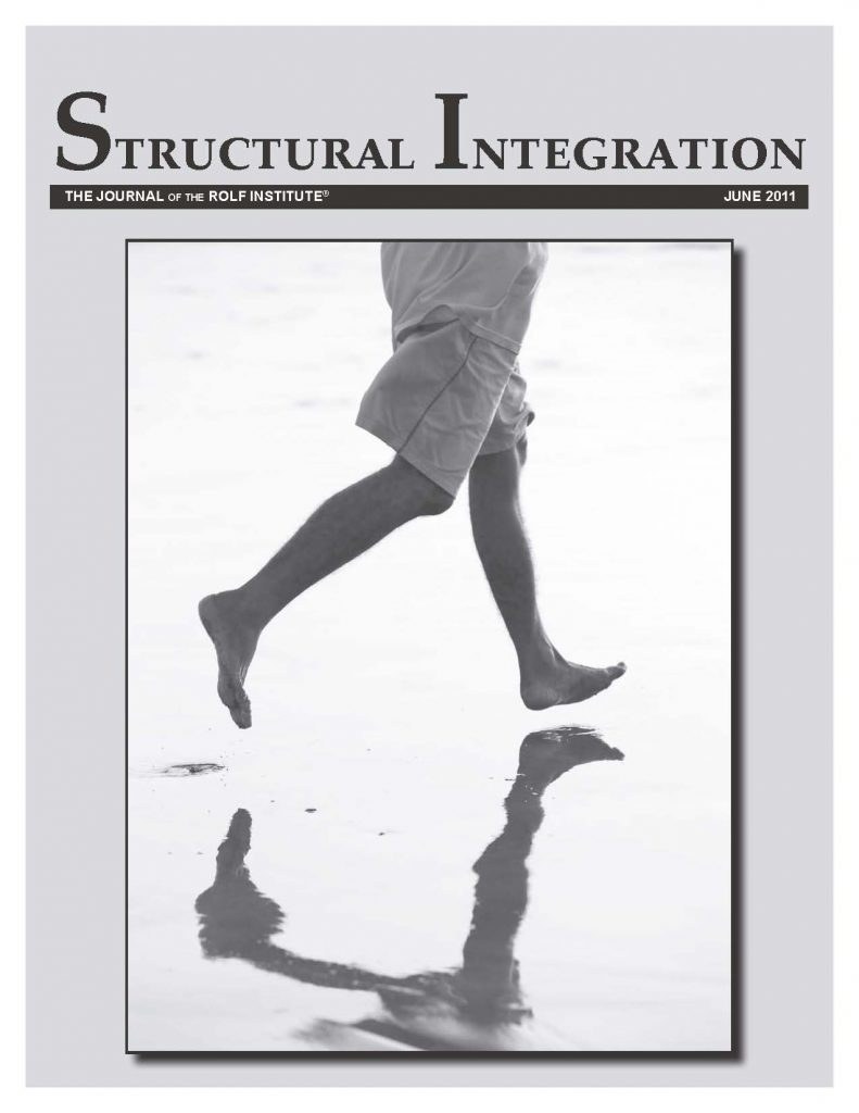
The human way of walking is strange. Not only does no other animal uses such a stiff stance leg, but no other animal moves its mass up and down so much with each step1 (see Figure 1). Generally speaking, to lift mass consumes energy, and no other animal voluntarily transforms flat land into hill country.
Moreover, a closer look shows that the upper body not only is lifted and lowered, but also changes its velocity in each step.

<i>Figure 1: An almost stiff stance leg moves the body mass up and down. In gait analysis, the “inverted pendulum” is the commonly accepted model for the basic pattern of human walking.</i>
Due to gravity’s action on the upper body mass, during the ascent the hip joint slows down, while during the descent it accelerates. To maintain balance, these changes in velocity must be transmitted to the upper body (see Figure 2). Alternately braking and pushing the combined mass of the trunk and head would also be expected to consume energy. In the convention of gait analysis, these two mechanisms – lifting and lowering, and braking and accelerating – are considered to consume considerable energy. Were our ancestors stupid to choose such a seemingly costly style of locomotion in a hot, arid environment?

<i>Figure 2: Besides the inverted pendulum of Figure 1 (the stance leg with its up and down), a second inverted pendulum exists – the upper body – which must be kept in balance although getting faster and slower all the time.</i>
Errors in the Conventional Model of Human Walking
Here we identify what we believe to be erroneous in the conventional model of human walking.
Consequently, to explain the “wasteful” muscle activity shown by EMG studies of walking, scientists like Hill and Fenn were seeking “chemical springs”, i.e., muscles that work “inversely” to produce chemical energy like accumulators when stretched eccentrically.7,8
The Role of Fascia
By accounting for the role of fascia, we can correct these errors and solve the puzzle. Let’s try a new hypothesis: that the lumbodorsal fascia, with a huge number of collagen fibers, acts as a tendon in counterpoint to the psoas major tendon, both of which are highly elastic and function as huge strong springs connecting the stance leg to the trunk (see Figure 3); and that these springs do most of the work of walking.

<i>Figure 3: A stance leg and a balancing upper body, connected by the lumbodorsal fascia and the gluteus maximus muscle.</i>
Some silly notions never die – and the nonelasticity of collagen is one of them. While the almost perfect elasticity of collagenous tissue has been proven repeatedly beyond any doubt and has been tacitly understood by many researchers, most physiologists still either deny outright that collagen is elastic or implicitly distinguish between collagen and “elastic fibers.”9 However, tendons and fascia actually consist of almost nothing but elastic collagen.
In our hypothesis, the HAT segment is role in walking. It functions as a heavy counterweight for the hip-extensor and -flexor springs. This would explain the human peculiarity of balancing a heavy weight high above the hip joint. Identifying springs in the upper body instead of in the legs also resolves the problem of the force directions.
While Fenn and many others considered absurd the possibility that in walking, tendons function as elastic springs held in tension by actively contracting muscles, it has been demonstrated as a reality for the gastrocnemius aponeurosis.10 The situation might be clarified if we can learn more about isometric muscular contraction, especially in the tonic, slow-twitch muscles. Surprisingly, there is virtually no research about the energy consumption of a muscle acting isometrically, far below maximum contraction force. If it turns out that this is not just a passenger, but plays an essential highly energy efficient, it will cast a whole new light onto endurance activities. And, it might also be that most muscles act this way in proper walking.
The Hypothesis of an Elastic “Bootstrap” Design
Have a look at Figure 4. In grey is the inverted pendulum of the conventional model of gait analysis. Superimposed on it in black is a representation of what our calculations have shown happens when the upper body is included. The stance leg moves up, thereby losing momentum and slowing down. Due to inertia, the HAT mass tends to maintain its speed. With no brake for the trunk, the walker would stumble forward. The opposite would happen when the stance leg goes back down: because of the weight it is supporting, the descending stance leg would accelerate while the trunk would stay behind, making the walker stumble backwards. As the conventional stance leg inverted pendulum does not work very well for the walker, it is evidently misleading.

<i>Figure 4: An upper body without extra support for balance (black) superimposed upon the conventional inverted pendulum (grey).</i>
Our hypothesis is that springs acting on the hip joint balance the weight of the upper body and avert the stumbling. To test our hypothesis, we developed the new model shown in Figure 5. This figure represents only the idealized single-stance phase of a step; the double-stance phase is not considered here. The springs counteract the inertia of the upper body, allowing it to maintain its balance on the alternately rising/decelerating and falling/accelerating hip joint base. At the same time, the springs pull the leg upwards in both phases, thereby also counteracting both the acceleration and deceleration themselves.

<i>Figure 5a: The first half of a step. A stance leg rises, due to kinetic energy and the action of the hip extension spring, which spring also decelerates the balancing upper body.
Figure 5b: The second half of a step. A stance leg falls, accelerated by the body weight and decelerated by the action of hip flexor spring, which spring also accelerates the balancing upper body.</i>
In our model, the inertia of the torso’s high and substantial mass is absolutely necessary as a counterweight for the pull of the spring. Observe also that this mechanism requires “horizontal” levers, which the shape of the human pelvis furnishes. What makes our model revolutionary is the absence of energy-consuming engines (contracting muscles). If it is congruent in its fundamental principles with reality, it explains a way of walking free of energetic cost. Because we are not aware of any theoretical discussion or empirical documentation of such a design in human biomechanics, engineering, or physics, we refrained from offering our hypothesis until we were absolutely certain of our calculations. At first, we doubted that it was possible to maintain upper body balance with no more than these two springs. However, our calculations show that when the spring tension is correct, the design works perfectly.
Not surprisingly, our calculations show that the success of the design depends on the precise adjustment of the springs, which would be achieved in the real world by fine-tuning the pre-tension of the relevant tissue structures. This tuning might be accomplished by muscles working isometrically under the control of accurate motor intelligence. As is the case with a fine musical instrument, effortless balance demands perfect harmony.
We consider our hypothesis to be a “bootstrap” design – a term derived from an 18th century German tale of one Baron Münchhausen, who reputedly pulled himself out of a swamp by his bootstraps. The same idea was introduced in English by James Joyce in Ulysses: The bootstrappers “had forced their way to the top from the lowest rung by the aid of their bootstraps. Sheer force of natural genius, that. With brains.” In engineering, a “bootstrap” is a device that feeds part of its output back into its input. Such devices are rarely so close to the original gravity-dependent meaning as ours.
The Anatomy of Elastic Walking
We propose that the combination of the lumbodorsal fascia and the gluteus maximus muscle serves as the hip erector spring (see Figure 6a) Indeed, EMG recordings of gluteus maximus activity support our hypothesis: maximum activity occurs at the beginning of the upward movement shortly after heel strike at maximum hip extension, when in our model the spring is stretched to its maximum length.11
Most likely, the psoas muscle and its long tendon serve as the hip flexor spring (Figure 6b). Unfortunately, little is known about the function of the psoas major in walking. Data on the “iliopsoas” almost always relates to the iliacus. Based on his observations of poliomyelitis victims, Duchenne12 concluded that the only muscles indispensable for walking, with or without mechanical support devices, are the hip flexors. Duchenne, by the way, because he could not use electrodes to stimulate the psoas itself, is responsible for popularizing the confusing term “iliopsoas.” Even when the function of the psoas major as a hip flexor is examined, its performance at an extended hip (Figure 6b) is often ignored (e.g., as in this source13).
In fact, the function of the psoas in walking is rarely examined at all. Most of the literature about the psoas addresses its assumed function as a stabilizer of both the hip joint and the lumbar spine (e.g. this source14). Bogduk disagrees. He reports, “A striking feature of the fascicles of psoas major is their similarity of length. This suggests that the psoas is designed to act from the lumbar spine on the femur. With all fascicles of similar length, they would all undergo the same relative shortening and would share to the same extent the linear excursion of their common site of attachment on the femur.”15 This seems to support our assumption that the psoas muscle acts mainly isometrically as an activator of the tendon spring.

<i>Figure 6a: The proposed hip erector spring – lumbodorsal fascia plus gluteus maximus muscle.
Figure 6b: The proposed hip erector and hip flexor springs – lumbodorsal fascia plus gluteus maximus and psoas major (tendon and muscle).</i>
Given the location of the psoas, EMG measurements in walking are rare. However, Keagy16 implanted electrodes into the psoas muscle during lumbar sympathectomy surgeries on five patients, and reported regular psoas activity in each patient during heel-rise. Guided by an ultrasound technique, Anderson17 inserted needle electrodes from the back in four subjects. At 1.5m/s walking speed, he measured activation duration times approximately 70ms while the muscle was elongating. Both studies support our hypothesis.
When the psoas is stretched most in walking, the hip is in internal rotation. “When through secondary muscle group action the hip is stabilized in a position of internal rotation, the action of the Iliopsoas is enhanced. At this time, the lesser trochanter is posterior and medial to the axis of the femur, and contraction of the Iliopsoas when riding anteriorly over the crest of the pubis produces reinforced and more deliberate lateral flexion and rotation of the spinal components through the transverse processes, which are posterior and lateral to the central axis of the vertebral bodies.”18 Because this rotation between pelvis and spine enhances the stretch of the lumbodorsal fascia on the other side, we speculate that the degree of rotation might indicate the degree to which both the psoas and lumbar fascia are utilized; i.e., minimal rotation might indicate low utilization of the psoas and lumbodorsal fascia, while the considerable rotations we have observed in walkers in remote Africa might indicate high utilization of those structures.
Practical Experiences
Because we speculate that improper or inadequate use of fascia might lead to back pain, we are experimenting with both Rolfing structural work and Rolf Movement education to help our clients use the fascia to achieve what we see as an elastic style of walking. It is difficult, given the subtlety of the adjustments, the need to involve the whole body, and the fact that “putting a spring into the step” seems to produce – or perhaps to demand – a change deep in the client’s personality.
If indeed the lumbodorsal fascia is rhythmically stretched during walking, we would expect it to produce a swing in the lumbar region (see Figure 7). Of course, this reminds us of Ida Rolf’s ideal of the “psoas walk.” We are now developing an optoelectronic device, with which we will be able to measure the swinging of the lumbar vertebrae. We have no choice but to succeed: the project is funded by the German government.

<i>Figure 7: A periodic stretching of the lumbodorsal fascia during walking should reveal itself by swinging lumbars.</i>
Our research has led us to a new and unexpected understanding of what “balance” actually means for the human body – an understanding reinforced by our practical experiments. The subtle adjustments to the springs before the start of each step are required in order to maintain not only spatial balance, but also temporal balance. This has compelled us to adopt a much more dynamic view of what human structure is about. As Rolfers, perhaps we should learn to perceive the specific appearance of elasticity in walking –i.e., the high-frequency oscillations at heel strike (and the resultant waves that ripple through the tissue) – as well as the appearance of harmonics and resonance. Perhaps we should be more attentive in general to the dynamic features of the body structure; as Bernstein observed as early as the 1930s, the human body structure is four-dimensional.19
<i>Zorn and Hodeck are physicists as well as Rolfers. Further information on this model is available online at www.swingwalker.net and in the upcoming book Dynamic Body, edited by Erik Dalton.
All images in this article are by the authors.</i>
Endnotes
Elastic Walking: The Fascial Engine[:]