

If you can see your path laid out in front of you step by step, you know it’s not your path.
Your own path you make with every step you take.Joseph Campbell
Have you ever heard the joyous shrieks of parents as their infant child takes her first steps? The “oohs,” “aws,” and digital recordings of these moments quickly become treasures in the familial archive. For an infant, rising-up to standing in the field of gravity is a feat of coordination and evolutionary impulse. However, the first steps taken by an infant are performed long before he or she rises to upright on baby feet. The gesture of alternate stepping begins in utero: prior to any kicking, stretching, or other motions an expectant mother feels pushing against her belly wall.
A developmental continuum exists between the first growth gestures and the later use of the legs. The activity or movement of growing shapes the means for the later activity of stepping. At every stage of its development, the embryo is performing expressive gestures out of which specific structures congeal. In fact, all patterns of behavior have embryonic developmental processes as their precursors.1
This article explores the slower growth and oscillatory movements of the embryonic limb buds, and specifically the hip joints and legs. These gestures are pre-exercised by the human embryo beginning at the end of the fourth week post-conception. These early formative patterns imprint the movement styles of the adult and are directly linked to some forms of hip dysplasia.
These movements define the formative pattering of the lower limb and occur at specific times in embryonic development. Imagine watching these movements in time-lapse sequence. We would see that the very growth of the embryonic lower limb mirrors the movement patterns necessary for walking.
Positional changes in the embryo’s limb anlage (precursor) can be detected as the limb grows in volume. These movements include growth-adduction toward the umbilical cord with a folding across the embryo’s genitofemoral fossa. This is followed by growth-flexion, the bend being the anlage of the knee. Subsequent embryonic events in the development of the lower limb include growth-extension of the knee, growth-flexion in a region that then becomes the ankle, and growth-eversion of the foot.2
Erich Blechschmidt, Ph.D., an embryologist, made serial dissections of embryos at every stage of development. He realized that genetics alone could not account for the precise formation and maturation of the fertilized egg. Instead, he cited the existence of metabolic fields of formation that shape the organism from outside to inside. The burgeoning field of epigenetics is developing an understanding of the external forces working in embryonic development. In other words, there are forces outside of the genetic markers activating gene expression. One of the ‘in-forming’ fields or kinetic blueprints is the ring torus3 (see Figure 1).
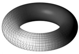
Figure 1: A torus (source: Wikipedia Media Commons).
Sensorial Inquiry: A Spiraling Torus
In the four-week-old embryo, the torus form shapes the caudal ring of ectodermal tissue that is pre-forming the pelvis, pelvic floor, and arising limb buds.
Oscillation: Streaming into Formation
Our limbs (and cortex) develop through slow oscillatory movements. Everything is vibrating with resonant intelligence. The body is movement. This shift in understanding challenges archaic beliefs that man is a machine and structure is inert. Mechanistic ideas about the body inhibit a leap into quantum understanding about the nature of physical reality, intelligence, and our transformational capacity. When thinking is sequestered by century-old ideas, it is easy to forget that all living systems emerge from oscillating and vibrating protoplasm. Our bodies are intelligence awakening. Most texts on embryological development depict static animations sequenced on linear timelines that omit reference to the continuum of gesturing life that informs and guides development. Embryonic growth is pulsing and alive; bursting forth like a garden in summer.
It was Blechschmidt who developed the concept of embryonic growth gestures, noting that we don’t have to remain fixated upon the static, lifeless forms of dissected embryos. “Nothing prevents our reckoning with the fact that the meticulously recorded positions and structures of the embryo are frozen records of what are actually developmental movements . . . . These movements are always more than just measurable changes of shape. They are always also the expression of living formations.4 [Italics added by this author for emphasis.]
The embryonic arm buds begin growing moments before lower-limb formation. The physical structures of adult arms and legs arise from identifiable gesturing. The future arms and legs of the embryo function in different capacities and their growth gestures mirror that difference. For example, limb-bud growth of the hands and arms displays a gesture of flexion and extension. Flexion speaks of a taking in or receiving while extension mimics giving or reaching out. The leg, led by the embryonic precursor of the paddle-shaped anlage of the foot, opens caudally from the oscillating torus of the lower body, pelvis, and lumbar plexus, with a functional gesture of stretching and extending.
In the early embryonic stages, a significant proportion of the leg’s inner tissue consists of nerve fascicles of the lumbar plexus. Thus it would appear that the nervous system is participating morphologically – and also functionally – in the growth-movements. “Given the improbability that any developmental process is exactly symmetrical, the growth stepping of one lower limb can be conceived as alternating with changes in position and form of the other limb. In this way, the cyclical patterns of fetal stepping are seen to be triggered by the embryo’s earlier growth-movements. On the other hand, the muscles arise initially as the passive elements of the limb’s musculoskeletal system, and it is only during late embryonic and early fetal stages that ‘spontaneous,’ more evident, patterns of muscular activity emerge.”5
Stephen Talbott writes that living organisms are gestured into existence, and are distinguished from other organisms by the character of this gesturing. With novel attitude, he suggests that this character does not disappear from the mature organism, but comes to expression at a different level. He reminds us that fixed form is always the end result of process and movement. Given our current habits of thought, we tend to start by conceptualizing already formed ‘things,’ which we then bring into movement or make into the causes of movement.6 Recognizing the origin of gestures as pre-forming structure inspires a shift in understanding and practice for both movement and body therapists.
We all believe it is we who move our own arms. And if we can catch within ourselves at least an inkling of that shaping inner gesture and impulse of will through which we bring our arms into outer movement . . . we may realize that the inner activity by which we move our arms is akin to the creative activity that first “gestured” the arms (or legs) into physical form.7
Sensorial Inquiry: Sensing the Oscillatory Patterns of Early Stepping
– The slow oscillation of a limb bud folding across the genitofemoral fossa toward the umbilical midline.
– The movement of bending, which pre-forms the knee.
– Growth-flexion in a region that pre-forms the ankle.
– Growth-eversion of the foot.
– Sense the continuum of these movements as you weave them together through your own body.
Hip Dysplasia
Rosemary Feitis and Louis Schultz write that we are embryos throughout our lifetimes. In their book The Endless Web, they show that the way the embryo-fetus lies in utero determines the ultimate pattern of the spine.8 This understanding applies to all aspects of formation. Hip dysplasia in adults can be traced to early limb-bud and bone development, or the lack of movement throughout a pregnancy (that is, the fetus’ ‘birth-lie pattern’; asymmetrical pressures and twists shape the embryo-fetus’s pliable body and mirror the mother’s alignment). Thus, the way a mother carries her body in pregnancy imprints the developing infant in utero. All of these factors and more may contribute to the malformation of the leg-acetabulum relationship.
Allow the following understanding to permeate your body-knowing: even before embryonic limb buds appear there are rotations, oscillations, and pulsations around a midline or longitudinal axis of orientation within the endocyst disc (the precursor to the embryo). “The surface growth of the ectoderm (a motor function) is already left-right asymmetrical. Perhaps an early event in limb asymmetry could be different surface growth rates in the ectoderm of the left and right.”9
The protoplasm of our beginnings is pulsating with the movement of life.
An understanding of embryology and the postnatal growth and development of the leg, hip, and pelvis contributes to a more complete understanding of hip dysplasia. This information can live in the back of your thinking mind, behind your study of adult anatomy and physiology, and it can inform your touch with the knowledge of the formative gestures shaping this the pelvic-hip-leg constellation.
Limb buds begin to appear in the fourth week post-conception. Hip formation begins in the seventh week. A cleft separates the tissue that will become the femoral head from the tissue that will become the pelvis. Through the oscillatory movements previously described, the femoral head begins to shape the cup-like recess of the acetabulum. If the head of the femur is not positioned properly in the acetabulum, or if movement of the femoral head is reduced, a shallow hip socket may develop. As mentioned previously, the intra-uterine environment also shapes this development.
By the eleventh week of gestation, hip formation is complete. However, at this moment of growth, the pelvis and femoral head are composed of cartilage that is soft and subject to the pressures and stresses imposed from the outer environment as well as within. Cartilage can be imprinted with these and other mechanical stresses, and the shape of the femoral head and acetabulum can be altered as well as the entire pelvic dynamic.
Oscillatory motion is implicit in formation. At twelve weeks post-conception, the lower limbs begin to rotate nearly 90° medially, so that the knees point anteriorly and the hips assume their normal position in the pelvis. Dislocations can occur at this time. Interference with the completion of this motion or the lack of rotation medially often occurs. In addition, as muscles develop they may exert varying pulls on the right alignment and positioning of the femur and acetabulum.
At birth, the hip joint is only partially developed, with less than 50% of the femoral head being covered by the acetabulum. The combination of these two factors can result in spontaneous subluxations and/ or dislocations from normal activity and kicking in some susceptible infants.
Embryological Understanding in Practice
When I first began studying embryology, I had no idea that it would profoundly affect my understanding of the human body. Initially, I felt detached from the pictures I studied in books or prepared for slide presentations for the classes I teach. With dedicated study and meditation, I began to understand the implications of what I was seeing and eventually sensing. Adult function and anatomy can be facilitated by studying the way our human form develops. In exploring this thread of our primal beginnings, embryology is a portal to witnessing the ‘origin of being,’ or consciousness arising and manifesting in the density of our form.
When an individual presents hip problems, gait problems, coordinative difficulties, or other distresses, I work with the formative patterning beneath the complaint. There is a prevailing belief in osteopathic medicine that the fields that generate the embryonic body continue to sustain our health and well-being throughout a lifetime. The embryo, as an archetype of perfect form and wholeness, serves as a blueprint for our body’s ability to heal itself. The formative and regenerative fluid forces that organize embryological development are present throughout our lifespan, available for harnessing of their therapeutic potency. In other words, the forces of embryogenesis become the forces of healing after birth.10 Through dedicated study and incorporating the perceptual understanding of a biodynamic practice, I have had the palpable sensory experience of the vast and interconnected matrix in which we are held. This matrix is a vital force of healing and is the resource for the transformational process we engage in with others and with ourselves. Appreciating the implications of embryology in our therapeutic practices may require a ‘quantum leaping’ beyond cherished belief systems, Newtonian mindsets, or linear space-time perceptions. The questions that I continually ask myself are: “What is a body?”, “What are the multiple dimensions that nourish and sustain physicality?”, and “Are we, as practitioners, able to open ourselves to the informational field in which we live?”
The linking back to the origin not only restores strength, but also creates the possibility of recognizing and bringing into play ever new chreodes (a biological pathway or habit) and new developmental lines.11
Carol Agneessens has been practicing Rolfing® Structural Integration for over thirty years and has been a member of the Rolf Institute faculty since 1993. Carol also offers trainings in Biodynamic Craniosacral Therapy. Visit her web site www.holographictouch.com for more information.
Endnotes
The Ground of Movement[:]