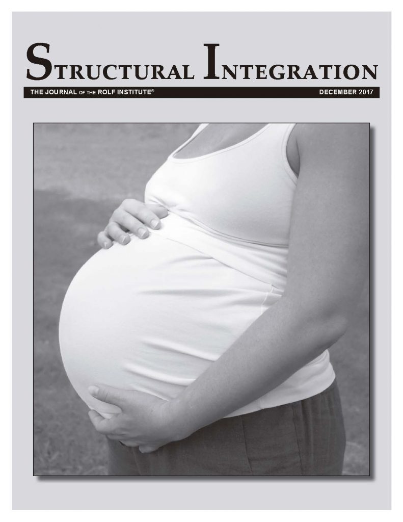
Introduction
All over the world, the legal and traditional regulations around pregnancy, birthing, and postnatal care are changing. I live and work in Germany, where, both by tradition but also by law since the 1930s, a midwife must be present at every delivery. Only in emergency cases are doctors alone allowed to help. Even in the case of a planned C-section, a midwife will be present in the operating room, firstly to take care of the woman, and then taking care of the baby. Frequently it will be the midwife, not the doctor, who carries out post-operative care. I received my training as a nurse and midwife at the university hospital in Würzburg, and this was the way I had worked for a decade when I started my Rolfing Structural Integration (SI) training almost twenty years ago. I witnessed about two hundred C-sections as the midwife in charge, and many more during my training and a one-month deployment in the operation ward.
Nowadays, in the US, Canada, and Europe, 23%-50% of deliveries are planned or end up as C-sections. Indeed, in more affluent socioeconomic groups in the US and South America, this percentage goes as high as 70%-90%. As the long-term effects and outcomes of C-sections become more apparent, authorities are now trying to encourage vaginal births. In Europe, the prevalence of birth by C-section is around 25%-40% and rising (some hospitals have a rate of 70%!), and equivalently there are increasing calls to return to more natural birth settings. But for the time being, we need to be able to work with the high number of women who have had a C-section.
Around 15% of women do not have children (depending on cultural and religious backgrounds, this figure may vary between 5% to 20%). Thus 85% of women have at least one child, and indeed many of them will have two or more children. This means that 30% to 70% of women who show up in our Rolfing practices have one or more C-sections in their history.
Many people think that the cesarean (or caesarean) section was named after Julius Caesar, the Roman emperor, and that he himself was born that way. But cesareans were actually already being performed (and called by that name) before he was born. The word comes from the Latin caedere: to cut. Women in those times did not survive a C-section. (Happily, we know that Caesar’s mother did live for many years after his birth, having given birth to him vaginally.) Abdominal cuts to save a child’s life were only performed when the mother’s life was already fading and could not be saved.
In the nineteenth century, rickets became very common due to malnutrition and the practice of keeping children indoors. It caused severe pelvic deformities, and many women died during labor at full term due to tearing of the uterine musculature. The medical alternatives then available were of little help: inducing labor prematurely might lead to death or handicap of the child, or the fetus might be killed in the womb; cesarean sections caused maternal death in 45%-60% of cases. In general, the mother’s life was saved over the child’s, and women were advised not to get pregnant again.
Only after the introduction of chloroform and antibiotics in the early twentieth century did C-sections become safer. Yet it is still the most common reason for maternal death in delivery (indeed, almost the only reason now in places like Germany). Today, the maternal mortality rate differs from 1:6600 live births in industrialized nations to as high as 1:250 births in underdeveloped / Global South countries. For the child, it also remains the case that a C-section is not the safest way to be born: breathing and blood-sugar regulation problems are just two possible adverse effects of being born by C-section. When these aspects are considered, it is possible that C-section threatens as many lives as it saves.
C-Section Procedure
Until the 1960s, the abdomen was cut lengthwise in a C-section, and the uterus opened transverse at the corpus; that is, at the cranial end. Sometimes even the uterus was cut open lengthwise. The standard method was formerly to cut all layers to the full extent of the opening required, and in the end suturing up every single layer one by one. Both ways caused large scars in the abdominal wall and, since the uterus was cut through the thickest part of its musculature, it would frequently rupture in later pregnancies and during labor. For this reason, it was recommended that after a C-section there should be no further pregnancies (requiring abstinence in those days). Later, with the advent of improved suturing, this advice was changed to: ‘once a C-section, always a C-section’, definitely the safer way to go.
From the 1960s onwards, surgeons began to prefer a suprapubic transversal cut of the abdominal wall (within the bikini zone), with a longitudinal cut of the muscular layer in the fascia, and then a supracervical transversal incision to the uterus. In this lower end of the uterus there is little musculature, and post-surgical ruptures are accordingly fewer. The materials used for suturing have improved considerably, and wounds heal much better, to the extent that a woman today might have a first child by C-section before going on to have three vaginal births.
The Misgav-Ladach method is also mainly used nowadays as it is considered less invasive and conducive to faster healing. In this method, it is only the outer skin layer that is cut all the way through; all the other layers are cut minimally (‘edgeless’) and are then opened by tearing or pulling. Only the uterus, abdominal fascia, and skin need to be sewn up afterwards; other layers (such as the layers of muscle and the peritoneum) are left open. The surgeon sometimes repositions these before suturing the next layer.
Preoperative Preparation
The following is standard preoperative preparation for a C-section:
Operation
A C-section operation consists of:
These steps are achieved with a lot of hurry and stress, because all of this needs to happen within one to two minutes from the start of the operation to avoid anesthetic and other medication getting into the child’s circulation. After the child has been separated from the mother’s circulation, the pace of the operation usually becomes more relaxed. Nevertheless, the operation continues to be potentially life-threatening for the woman.
The baby’s vital signs will be checked; in the past, the child would be taken away to the neonatal ward (nursery) and brought to the woman maybe a day later, or even days later, when the mother was able to walk over to the nursery. Nowadays, the baby will be checked and brought to the mother straightaway and placed on her upper thorax so she can see and feel the baby. Only later will the baby be brought to the nursery for a complete check-up. If the baby has problems, the mother might not see the baby until she is able to walk or be brought to the neonatal intensive-care unit.
The operation continues:
Postoperative Care
Next we have the postoperative care procedures:
Recovery
While all these steps are carried out for good reasons, many of them may have long-term effects on the body. In pregnancy and delivery, the body’s own capacity to heal and reposition itself in space and gravity is abundant. (One example makes this brilliantly clear: surgeons used to combine some C-sections with sterilizations, but stopped doing this after they found that – compared with the usual refertilization rate of 0.1% – sterilizations carried out in the two weeks after birth have a refertilization rate of up to 10%.)
Nevertheless, the woman’s body and being have a lot to do (not forgetting childcare!) after an operation as major as this. How well a woman heals after a C-section will depend on a number of factors, including her physical state prior to the birth and the resources available to her after the operation. It may be easier to recover from a C-section that was planned, but this also depends on whether her expectations of recovery were realistic. For women who chose to give birth vaginally, but found themselves overrun by emergency and hospital procedures, there will be a need to process the psychological themes of their experience alongside the physical effects (and all of this while caring for a newborn). This can be absolutely overwhelming. A great deal of tender and empathetic support is needed for many weeks after the operation.
There are five typical stages of recovery:
Phase One: In the first few hours, immediate survival is in the foreground. There may be shocked numbness. Basic body functions are important; thinking may be too demanding.
Phase Two: In the first few days, the numbness dissipates, and the focus shifts to pain management, relearning to walk, re-engaging bladder control, and the resumption or normal bodily functions. Managing pain around daily activities will be a challenge, and emotions start to show up (sorrow, disappointment, anger, and feelings of guilt may be among these). The typical ‘baby blues’ often marked by excessive crying may emerge at around the third day.
Phase Three: Awareness. The period after getting home and until around eight weeks after delivery is a hard time for all women, especially after a C-section. The round-the-clock care needed by a newborn inhibits a normal recovery from a big operation. Doubts, recriminations, and judgement (by the woman or by others) around the decision to have a C-section, or the question of how it might have been avoided, are foreground. For many, this can be a very emotional and intense phase, as women try to negotiate whether they or their medical team were at ‘fault’.
Phase Four: Medium-term resolution. In the period two to twelve months after the birth, women try to make sense of their experience. They may recapitulate what happened from different aspects, and try to settle on the facts. This is often the time when they begin to seek help with the outcomes of their experience (meeting Rolfers and other practitioners, restarting exercise, finding groups to talk about their experience, etc.). Some women may nevertheless suppress the reality of the operation and try to get back to a body image from before the pregnancy.
Phase Five: Resolution. Acceptance and allowing the experience to be an integrated part of their lives.
Let’s go back to the physical part of the C-section. I will point out common effects of many of the steps listed above. Of course, this list is not, and could never be, complete, and will not be true of every woman you meet in this situation. Women may experience one or several of these side effects, and may not be able to name or give words to some aspects of their physical state. The following is presented as a prompt to help you better identify, understand, and evaluate your clinical observations.
General Themes for a Rolfing Body Reading
Based on what has been discussed above, there are many aspects Rolfers can consider in the structural and physical assessment of a client who has undergone a C-section, depending on their scope of training and other licensure. Some of these include:
Common Reactions to the C-Section Procedure
In general, pregnancy hormones result in tissue that is more elastic and less able to recoil. This means that dislocation can occur more easily and be harder to heal. These hormones are active for several weeks after delivery and throughout breastfeeding. While it is easy to affect the body through Rolfing SI during this time, it is also easier to dislocate joints or overstretch tissues, and beneficial effects may not be lasting.
Anaesthesia used during a C-section may be local or general. With local anesthesia the lower part of the body will be numb, while in general anesthesia the patient will not feel dislocations and strains to joints and tissues. (Of course, this is true for every operation, not only C-sections.)
The intravenous drip is usually placed on the right arm, and this arm will be fixed in place during the operation so that any unconscious or inadvertent movements will not tear out the access to the blood system. The arm will be held in place for the duration of the operation (thirty-five to sixty minutes on average). Very often, the arm is not fully comfortable during the operation, and this can lead to slight dislocations, ligamentous overstretching, and muscle contractions that may afterwards cause inhibitions in the right shoulder. The oxygen sensor is placed on one finger on the same side.
Blood-pressure measurement is done on the other arm. The sphygmomanometer stays in place for the whole operation and for some hours afterwards, and can cause inhibited movement in the arm and shoulder.
During the operation, the body is fixed on the operating table in lithotomy/dorsosacral position (on the back with flexed hips and knees and legs splayed), and also in the Trendelenburg position, meaning slightly turned to the left, to avoid vena cava syndrome. Reclining fixedly in this position for more than half an hour, with a twisted sacrum, can distort the whole lower spine and especially the sacroiliac joints (if there is preexisting misalignment, you can imagine the effect). The surgeon will be located on the left side of the body, with a helper on the right side. Another medical assistant may also be standing at the end of the surgical table, between the patient’s legs. With general anesthetic, the neck may be overstretched, and vertebrae may become fixated or misaligned.
After the operation, the woman will be placed in a regular hospital bed, but this may be achieved a little carelessly. As it is hard to move one’s own body immediately after an operation, all sorts of ‘minor’ misalignments may occur.
Over the coming days, patients are mobilized quickly, with a view to discharging them from the hospital and allowing them to be at home with their new baby. However, this is achieved with a lot of painkillers that enable the woman to get out of bed at a time when she should still be in recovery. Not infrequently, women experience secondary infections due to wounds reopening, and may even need another operation to clean out infected cavities or remove excessive scar tissue and adhesions.
Other Reactions
There are other reactions to treatment that are often neglected or not immediately obvious.
How the Ten Series Supports Recovery from a C-section
There are many ways a Rolfing Ten Series can support a woman in recovery from the effects of a C-section. If you are in any doubt that a client’s presentation is within a normal range, do not hesitate to refer her to a physician prior to working with her.
Session One can help address the effects of both pregnancy and delivery on the breath. During pregnancy, the lower ribs are pushed outwards to make space for the baby, with pregnancy hormones working to soften ligamentous structures. This may mean that the ribs are not in place, or that the diaphragm needs to return to normal function. During delivery (and especially an attempted vaginal delivery), the breath may have been used to try to push the baby out.
Session Two can address the effects of pregnancy on plantar support, as well as the mobilization under stress of trying to ‘kick’ a perceived aggressor or ‘run away’ from the situation in the hospital.
Session Three can resolve issues in front-to-back balance that may be due to internal strains on the pubic symphysis and dysfunction of the lower abdomen.
Session Four can help with irritations to the pelvic floor created by internal pulls, and with squeezing the pelvic floor due to trauma, numbness, and feared or real incontinence.
Session Five can really help with finding space for the abdominal organs and psoas, as well as with issues with both body schema and body image with respect to the belly.
Session Six can address issues around sacral dislocation and the effects of internal pulls on the mesentery.
Session Seven can resolve issues from the positioning of arms, shoulders, and neck during the operation.
Sessions Eight to Ten, the final three sessions of the Series, help to re-establish fascial lines and transitions throughout the whole body, and help the client to address issues with both body schema and body image.
Conclusion
I hope that this article can contribute to a better understanding of the aftermath of C-sections and help us, as Rolfers, to resolve the difficulties they create for our clients. The Rolfing Ten Series is wonderfully designed, and applied carefully it may help with many of the effects of a C-section.
Dorit Schatz trained as a nurse and midwife in Würzburg, Germany, completing her training by state examination in 1991. She worked in hospitals for ten years, spending her spare time training in shiatsu, craniosacral therapy, reflexology, homeopathy, and other modalities. In 1998, she left the hospital to complete her Rolfing training in Munich. Dorit completed her Advanced Rolfing Training in Santa Monica in 2002, and her Rolf Movement Training in Munich, also in 2002. Dorit has assisted several Basic Rolfing Trainings, and was a participant in the European teacher training. She lives and works in Munich. In her practice she mainly works with ‘regular’ Rolfing clients, but additionally specializes in themes around pregnancy, postpartum, and pelvic-floor issues.
Bibliography
Anzieu, D. 1996. Das Haut-Ich. Frankfurt: Suhrkamp Verlag.
Barral, J.-P. 2004. Viszerale Osteopathie in der Gynäkologie: Urogenitale Manipulation. München: Elsevier. [Available in English under the title Urogenital Manipulation (Eastland Press, 1993).
Bordier, G. 1980. Anatomie Appliquée à la Danse: Le corps humain instrument de la danse. Paris: Amphora.
Calais-Germain, B. 1999. Le Perinée Feminin et l’accouchement – Éléments d’anatomie, Applications pratiques. Micropolis: éditions DesIris.
Debroux, J.-J. 2004. Faszienbehandlung in der Osteopathie. Stuttgart: Hippokrates.
De Jong, T. and G. Kemmler 1996. Kaiserschnitt, Narben an Bauch und Seele: Ein Ratgeber für Kaiserschnittmütter. Frankfurt: Fischer.
Pschyrembel, W. and J. Dudenhausen 1994. Praktische Geburtshilfe mit geburtshilflichen Operationen. Berlin: De Gruyter.
Hees, H. and F. Sinowatz 2001. Allgemeine und spezielle Pathologie. Köln: Deutscher Ärzteverlag.
Hertling, D. and R. Kessler 1983. Management of Common Musculoskeletal Disorders: Physical Therapy Principles and Methods. New York: Harper & Row.
Jelkmann, W. and F. Sinowatz 1996. Physiologie. Köln: Deutscher Ärzteverlag.
Martius, J. 1998. Hebammenlehrbuch. Stuttgart: Thieme.
Martius, J. and A. Nowotny 1969/2006. Gynäkologie, Geburtshilfe, Neonatologie: Lehrbuch für Pflegeberufe. Stuttgart: Kohlhammer.
Meert, G. 2006. Das Becken aus osteopathischer Sicht: Funktionelle Zusammenhänge nach dem Tensegrity-Modell. München: Urban & Fischer.
Odent, M. 2014. Generation Kaiserschnitt: Wie die moderne Geburtspraxis die Menschheit verändert. München: Random House.
Schultz, R. 1999. Out in the Open: The Complete Male Pelvis. Berkeley, California: North Atlantic Books.
Tanzberger, R., A. Kuhn, G. Möbs, and U. Baumgartner 2013. Der Beckenboden. München: Urban & Fischer Verlag.
Tilscher, H. 2005. Die Wirbelsäule der Frau. Vienna: Verlagshaus der Ärzte.Rolfing® SI After Cesarean Section[:]
To have full access to the content of this article you need to be registered on the site. Sign up or Register.