
Read books and study nature.
When the two don’t agree,
Throw out the books.
– William Albrecht, PhD (1888-1974),
renowned soil scientist.
(Note that this quote is considered a paraphrase from his writings rather than a verbatim passage, often attributed to his 1975 book, Albrecht’s Foundation Concepts.)
Where This Story of Forces and Stressors Begins
My mountain bike tires slipped on a wet clay berm. As I was thrown off my bike, I attempted to run it out, but my foot collapsed beneath me. When I tried to stand up, I realized that my right foot was hanging off the tibia, slightly supinated and rotated externally. Thankfully, there was no blood. I yelled to my friends behind me, “I’m down, and we’re going to the emergency room for surgery!”

my leg or while they half-carried me and I half-scooted on my butt out of the forest towards the logging road where the ambulance picked me up. In fact, I spent most of my time in the ambulance and the emergency room, cracking jokes and sharing stories with the paramedics, nurses, and doctors.
Hypermobility means different things to different people.
For me, it means that when I was younger, I used to do a lot of fun party tricks like walking with backward knees mimicking a Barbie doll. When I was thirteen, my family doctor told me that I would one day struggle with pregnancy because of my lumbar lordosis. I heard him, but he didn’t identify what I could do to change it.
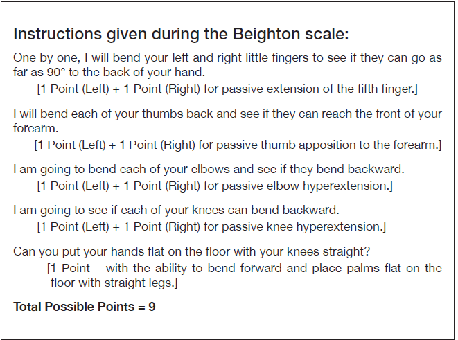
Using the Beighton score for hypermobility, I scored 7 out of 9 at an adult age when many have lost their flexibility (see Figure 1). But I never knew that hypermobility was diagnosable until this foot injury.
Dr. Tina Wang and colleagues (2025) state that those with hypermobility Ehlers-Danlos syndrome (hEDS) and hypermobility spectrum disorders (HSD) often have joint hypermobility and instability, skin hyperextendibility, and connective tissue fragility. People who have hEDS/HSD struggle with having a fascial system that includes a dysregulated extracellular matrix including disorganization of type I, III, and V collagen. And they have altered fibroblast-to-myoblast activation in dermal tissue and the deep fascia. This leads to denser tissue in some areas, lax tissue in other areas, chronic low-grade inflammation, and pain. In addition, the myofibroblasts remain in the tissue longer than normal when the cellular repair mechanisms are activated during wound healing and fibrosis. Ultrasound studies demonstrate that those with hEDS/HSD have altered deep fascia of the sternocleidomastoid, iliotibial tract, thoracolumbar fascia, and fascia iliaca.
Prior to this injury, I didn’t feel much pain or discomfort unless I stretched too far. Then the pain felt ‘nervy’, and I was unable to sleep until I helped the nerve find its way home again. I have to be cautious about who works on me and the type of bodywork I get. More than twenty years
______________________________________________________________________________________________________________________________________________________
ago, before I found Rolfing® Structural Integration, the pain was systemic in my knee, hip, head, neck, and left ankle. I was a wreck with very flexible hip, knee, and ankle joints, while my lumbar, thoracic, and back of the neck fascia felt dense and twisted.
As Rolfers™ know, and as Dr. Wang and colleagues have described (2025), fascia is important for structural support, proprioception, systemic communication, and mechano-transduction. Not only did Rolfers and Rolfing students help smooth and untwist my fascia in 2005 when I went through my Rolfing training, at the time I was a recovering twisted snowboarder, they also helped me become embodied, to pay attention to the gentle, quiet pain signals before they became screams.
Yet, I don’t think any of us recognized my hypermobility as an issue, although I would beg practitioners to not pick up my foot when I was laying supine, because I don’t like the feeling of my knee hyperextending in space.
So, why don’t I feel much pain? I attribute my low pain level to my diet, receiving many hours of Rolfing® sessions, the fascia work I do on myself regularly, and my movement practice that includes daily stretching and loading my joints while carrying heavy loads like hay bales and water buckets, and while enjoying hiking and downhill skiing. But a low level of pain does not mean someone is structurally balanced or functioning well. It just means the pain hasn’t hit yet.
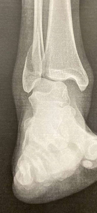
When the emergency room osteopath showed me the X-ray and pointed out the broken fibula, which was displaced, and the very large, clear medial space between my talus and tibia, all I could think was that my road to recovery would be a long one. The clear medial space, which is a radiographic measurement to diagnose ankle fractures, is considered too large if it is above 4 millimeters. Mine was 13 millimeters. He didn’t mention that every toe, metatarsal, and tarsal was rotated and/or translated. But I could see the rotations in the X-ray and my big toe, which peeked out from the splint. The most the DO could do for me was to apply traction to my fibula, put me in a cast, and recommend surgery.
The most discomfort I felt was at my medial meniscus of my right knee, which I feared would tear with the weight of the cast and the rotations of my femur and tibia. When I looked in the mirror, I could see that not only did I break my fibula, but my femur was adducted and medially rotated. Thankfully, Advanced Rolfer Jon Martine had taught me how to gently rotate my medial meniscus when I had strained my anterior cruciate ligament in 2005. So, while I sat on the couch for twelve days waiting for ankle surgery, I did what I could for the rotations of my pelvis, femur, and medial meniscus.
Having Surgery While Hypermobile
At the pre-operation appointment, the surgeon suspected that I had completely torn my deltoid ligament, along with breaking the fibula, and tearing the syndesmosis, the fibrous joint binding the distal tibia and fibula together by a strong sheet of connective tissue. I begged him to be careful with my easily hyperextendable knees during surgery. But he didn’t seem concerned when he determined that I don’t have hEDS. Before the surgery, my pain level remained at a two. Although I was uncomfortable sitting and lying down for so many hours.
Today, both hEDS and HSD are considered multisystemic connective tissue disorders featuring not just musculoskeletal issues but also including immune system dysregulation, chronic pain, low-grade inflammation, as well as visceral and nervous system dysfunction. Now we know, fascia is everywhere! Those with hEDS/HSD also tend towards proprioception deficits and frequent injuries.
Interestingly, my husband and younger son are also hypermobile. My older son, who is adopted, has no hypermobility. He has never been able to touch his toes. Although we are all athletic, my husband, younger son, and I all fall a lot while skiing. My oldest son rarely falls at all. And while playing soccer, my youngest son falls more than any of his teammates, even though he is quick and agile. And he sprains his ankles far more than his teammates.
The last thing I said to the surgeon and his team before the anesthesia knocked me out was, “Please, please, please be careful not to hyperextend my knees.”
The surgeon screwed a plate along my distal fibula with seven screws and

attached a button to my tibia to anchor my deltoid ligament from the calcaneus to the tibia. Then a cord was attached from my deltoid ligament to the button on my tibia, then pulled across to the fibula. Because my lower leg was still unstable, he also screwed my fibula to my tibia to allow the syndesmosis to heal.
Three days after the surgery, the pain level lowered from a six to a three, but a dull nerve ache began in my calcaneus, especially when I slept on my back. It felt as if someone was pushing my calcaneus and tibia posterior into the bed or sofa. And my knee felt chronically hyperextended. It was the strangest sensation, which often kept me up at night. My sense is that during the surgery, the combination of anesthesia and hypermobility made my lax ligaments even looser, and the tibia and fibula shifted posteriorly relative to my talus and calcaneus. Then the surgeon screwed me back together.
Surgical Recovery in a Hypermobile Body
Six weeks later, when I was cleared for a walking boot and physical therapy, my work of untwisting the bones of my foot, leg, and pelvis began. Because I am so flexible, the physical therapists assumed that I would regain function quickly, and were obviously surprised when I didn’t. The hamstring and calf stretches they suggested to increase dorsiflexion were no help. Eight weeks after the boot came off, plantarflexion remained extremely difficult, causing my foot to supinate into a kidney bean shape. Dorsiflexion also remained limited at 8 degrees, whereas I had about 12 to 15 degrees on my unaffected leg.
All along the metal plate, along the periosteum of my tibia and fibula, and along my Achilles, especially by the calcaneus, the fascia wouldn’t release easily. This made me think that the injury and surgery led to increased fibrosity, where the fibroblasts transitioned to myofibroblasts, creating densification in the whole region (Wang et al. 2025).
In bed, I had to wrap a cashmere scarf around my lower leg and knee, then tuck the ends of the scarf into a sock near the heel to keep my tibia from dropping posterior relative to my talus. I did a lot of taping the ankle at night, as if I had a lateral sprain, and it helped a bit, but not as much as the scarf.
Ida P. Rolf, PhD (1896-1979) wrote, “The bones of the legs have to be directly above the ankles” (1978, 97). Since the accident, my right leg bones are not. They are behind the ankle. With every step I took after the surgery, especially in the morning, it felt like my weight landed on my heel rather than mid-foot.
When a physical therapist tractioned my foot distally, attempting to release the fascia and give my ankle joint more space for the talus to move, it felt like they were detaching my foot and stretching it into a clown foot. This type of traction probably works well for most. But it made me sick to my stomach. I think this is due to a combination of the hypermobility and the traction when they pulled on the foot in the emergency room and during the surgery.
Strangely enough, although my tibia felt posterior and my knee felt hyperextended while lying flat, when upright in gravity, I was unable to straighten my knee. It wasn’t until I wrapped my hands around my calf and slacked my leg toward the hip

socket, and rotated my femur externally, that I began to feel a stretch in my calf at the ankle. Between slacking the tension of the leg, general and thorough fascia release, and cupping the dense tissue at my Achilles and along the fibula, eventually the fascia along my lower leg began to lengthen and become more organized. Nowadays, when I exercise or do my stretches, I imagine that the muscles and tendinous slings strengthen and contract upwards to keep my tibia and fibula from collapsing onto my talus. At six months post-surgery, I still could not dorsiflex or plantar flex enough to run. Part of the issue may be that the screw between my tibia and fibula is limiting rotation. With every step I take, especially during push off, I often feel the strain around the cuboid, fourth and fifth metatarsals, the talus, the metal plate, and the bone break. My foot often curves into the kidney bean shape and stresses the bone break if I haven’t stretched my adductors and calves enough. In one of my notebooks from an annual meeting, Advanced Rolfing Instructor Jan Sultan said something to the effect of, “In order to have a functioning transverse arch, the lateral arch needs to function well.” And the structure of my lateral and medial arch is compromised, which means I can’t push off well, and I lack spring in my step.
The Advantages of Being a Rolfer™
If I didn’t have my Rolfing skills, I don’t know what I would have done. Every morning, in order to walk with minimal pain, I have to push my tibia and fibula anteriorly, rotate the fourth and fifth metatarsals and cuboid internally while translating them medially, and slack my tibia and fibula superiorly and medially. As time goes on, I can feel my connective tissues becoming stronger and holding the bones in place better. But from prior injury experience, it will be about a year before I won’t have to work on my foot daily. Strangely enough, the most helpful therapy has been skiing. Once I could get into a ski boot, I was forced to lean forward. My Achilles stretches in this position and it feels less dense, longer, and less interwoven with the fascia around my ankle.
Rolfers, We Deal with the Unexpected When I have shown the rotations in my ankle and foot to the foot-specialist physical therapists, they appear confused. Osteopathic books don’t seem to address the twists I see in my foot. And this is why I love Rolfing® Structural Integration! Because it taught me to trust my eyes and hands, even if they don’t match what books and experts say. Rolf wrote, “Our job is to learn to contact reality as the reality exists in the human body. Not to contact names, not to know the names, not to know abstract patterns” (1978, 206). “The whole trick of Rolfing [Structural Integration] is going around and around and around, and not barging through” (1978, 83). “Fascia is the organ of posture” (1978, 124). When I come up for air from working on my foot and ankle, I remind myself that because fascia connects to everything, to look above, look to the other side, and to think about how the accident affected the organs. The work on my trochanter helps my ankle and foot. The work on my third and fourth lumbar vertebrae helps the ankle and foot. Stretching my adductors helps the ankle and foot. Strengthening my core in the bridge yoga pose helps the ankle and foot. While focusing on the upper segments of my body, I self-slack, supporting my long bones towards the acetabulum and sacrum. This limits how much they collapse onto the talus. Moshé Feldenkrais (1904-1984), founder of the Feldenkrais method, wrote, “In order to change our mode of action, we must change the image of ourselves that we carry within us” (1980, 10). I find that as my core becomes stronger from the daily yoga bridges that reduce my lumbar lordosis, my image of myself as a floppy person is changing. I am standing taller. My quadriceps, which have been strong and contracted from pregnancy and years of skiing, biking, and sitting, are lengthening, as is my psoas. It’s a different kind of taller than what I feel while experiencing Rolfing sessions, or even with stretching and yoga, because I am strengthening my weaknesses, which are partially due to my lifelong hypermobility. Who knows how much the loss of knee extension, ankle dorsiflexion, and plantar flexion is due to the twists in my foot, pelvis, femur, tibia, and fibula, or due to the hardware, or due to hypermobility? This is what I know: I will never stop learning and working for fluid movement in gravity, despite hypermobility and hardware.
Author’s note: All my thanks go to my Rolfing instructors and colleagues. I am so grateful that I found this career and this way of looking at the human body. It saved my life once and my mobility many times. This article was inspired by Rolfing Instructor Valerie Berg’s March 2020 article, “The Complexity and Reasoning of Keeping 3D Hands and Fingers,” about healing her arm after a car accident, and science journalist James Nestor, and his 2020 book, Breath: The New Science of a Lost Art. I love it when writers investigate their own healing process.
Luca Williams is a Certified Rolfer® and newly Certified Functional Medicine Nutritionist, who will one day become a Certified Advanced Rolfer when she’s ready to leave her family for six weeks. Her website is lucasrolfing.com. She also writes fiction with a Panamanian-American magical realism angle under the name of Luca Brite. See lucabrite.com.
References
Albrecht, William A. 1975. Albrecht’s foundation concepts: The Albrecht papers, volume I. Viroqua, WI: Acres U.S.A.
Berg, Valerie. 2020. The complexity and reasoning of keeping 3D hands and fingers. Structure, Function, Integration 48(1):20-28.
Feldenkrais, Moshé. 1980. Awareness through movement. London, UK: Penguin.
Nester, James. 2020. Breath: The new science of a lost art. New York, NY: Riverhead Books.
Physiopedia contributors. 2025. Beighton score. Physiopedia Available from: https://www.physio-pedia.com/index.php?title=Beighton_Score&oldid=365776.
Rolf, Ida P. 1978. Ida Rolf talks about Rolfing and physical reality. (R. Feitis, ed.) New York: Harper and Row.
Wang, Tina J., Stecco, Antonio, Hakim, Alan J., and Robert Schleip. 2025. Fascial pathophysiology in hypermobility spectrum disorders and hypermobile Ehlers-Danlos Syndrome: A review of emerging evidence. International Journal of Molecular Sciences 26(12), 5587.
Keywords hypermobility; hypermobility spectrum disorder (HSD); Ehlers-Danlos syndrome (hEDS); Rolfing Structural Integration; fascia; ankle fracture; fibula deltoid ligament; syndesmosis; structural recovery; ankle; fascial densification. ■