

Authors’ note: This article is in two parts: Part 1 is written by Lina Amy Hack, edited by Dr. Stephen W. Porges, and generously contains the 2025 Fascia Research keynote slides and notes of Dr. Porges. Part 2 is an edited conversation recorded on February 19, 2026, over Zoom. The interview has been condensed and lightly edited by both authors.
Part 1: Polyvagal Theory, the Autonomic Nervous System, and Fascia “Polyvagal Theory highlights the crucial role of the body and nervous system in health and well-being, demonstrating how our thoughts, emotions, and behaviors are shaped by our body’s ongoing assessment of our level of safety or threat. It underscores the bidirectional communication between the body and the brain and the necessity of fostering safety and connection to enhance overall health.”– In Safe and Sound: A Polyvagal Approach for Connection, Change, and Healing (Porges and Onderko 2025; 2). A Foreword by Lina Amy Hack I came to this work as a curious student, not an expert. What follows are my best class notes after more than twenty years of reading Dr. Porges’ research, books, and attending his lectures. This summary is the information wish I could have had the first time someone said to me – have you heard of Polyvagal Theory? Better yet, Dr. Porges himself edited this essay, and gracefully wove correctness into the information. It’s a honor to have my favorite professor edit my work to make it an A+ paper. Enjoy!
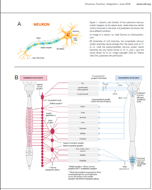
Polyvagal Theory proposes that the vagus nerve is not a unitary structure conveying a single “rest and digest” signal, but rather a complex system composed of multiple functionally distinct pathways. Although traditionally described as one nerve, the tenth cranial nerve (CN X), the vagus is a paired, bilateral nerve, with left and right branches emerging from corresponding regions of the brainstem. It is a major component of the parasympathetic branch of the autonomic nervous system (ANS), yet its function extends beyond a simple efferent role (see Figure 1), more than neural signals from the brain to the body. Historically, the vagus nerve was conceptualized as a singular pathway transmitting a generalized inhibitory signal from the brainstem to visceral organs. The vagal signal slows down the heart rhythm, for example. This view is now recognized as overly simplistic. Approximately 80% of vagal fibers are afferent, conveying sensory information from visceral organs to brainstem structures (Porges 1995, 2007, 2023). Thus, the vagus primarily serves as a bidirectional communication pathway, with a dominant role in transmitting information about the physiological state of the body to brainstem foundational circuits. The efferent (motor) component of the vagus, comprising roughly 20% of its fibers, is not functionally uniform. Polyvagal Theory distinguishes between two primary vagal pathways originating
in different brainstem nuclei: a ventral vagal pathway, arising from the nucleus ambiguus, and a dorsal vagal pathway, originating in the dorsal motor nucleus of the vagus. These pathways differ in their phylogenetic history, neuroanatomical organization, and functional roles in regulating physiological state and behavior.
A Brief Note about the ANS
“The ANS is a brain-body network that coordinates metabolic processes to support organ function and promotes biobehavioral reactions to danger and anticipated threats” (Jokić et al. 2023, 160). The ANS, and the role of the vagus nerve within it, are complex and often oversimplified. Neurons are the fundamental cellular unit of the ANS. Their cell bodies cluster to form nuclei – regions of grey matter located within the forebrain, brainstem, and spinal cord – as well as ganglia in the peripheral nervous system. Peripheral ganglia often appear, in dissection, as bead-like structures connected by neural tracts, forming paired chains along the anterior aspect of the spinal column. Axons bundle into nerve pathways that transmit signals over varying distances, from short projections between adjacent brainstem structures to long pathways such as those linking visceral organs (e.g., pancreas) to brainstem regulatory centers via the vagus nerve. Functionally, the ANS has two primary peripheral divisions: the sympathetic and parasympathetic divisions. Sympathetic neurons originate in the thoracolumbar spinal cord (T1 to L2) and project through the sympathetic chain ganglion to target organs, including the heart, lungs, vasculature, and gastrointestinal tract. These pathways support mobilization – classically described as “fight-or-flight” – enabling rapid physiological responses to challenge. Sympathetic activation is coordinated with central regulatory structures, particularly the hypothalamus, which engages neuroendocrine pathways. Under conditions of threat, hypothalamic activation can initiate the hypothalamicpituitary- adrenal (HPA) axis, resulting in the release of glucocorticoids and catecholamines that support sustained metabolic mobilization. In other words, the adrenaline rush. Parasympathetic neurons originate in brainstem nuclei and the sacral spinal cord. Cranial parasympathetic outflow is conveyed through cranial nerves: – three (CN III; oculomotor nerve), – seven (CN VII; facial nerve), – nine (CN IX; glossopharyngeal), – and ten (CN X; vagus nerve), while sacral pathways (S2 to S4) regulate pelvic organs. Traditionally, the parasympathetic system has been characterized as promoting the “rest-anddigest” functions and conceptualized as antagonistic to sympathetic activation, when one is up, the other is down, and vice versa However, this binary framework is insufficient. Polyvagal Theory (Porges 1995, 2007, 2023, Porges and Porges 2023) reframes the parasympathetic system – particularly the vagus nerve – as comprising multiple pathways with distinct evolutionary origins and functional roles in regulating physiological state, behavior, and social engagement.
From 2005 to 2015 to 2025
My first exposure to Polyvagal Theory occurred in 2005 during a Somatic Experiencing® (SE) training in Canada. The instructor emphasized that, in response to life threat, the body does more than mobilize through fight or flight; it can also engage an immobilization response. At that time, the concept of a “freeze” response was not widely recognized as an adaptive biological strategy. Individuals who became immobilized during a threat
– Neurons are the base cellular unit of the ANS.
– The location of the cell bodies reflects function; they are called nuclei.
– The path and destination of the emerging axons determine where the signal is delivered, from the cell body to the terminus of the axonal tract.
– Hypothalamus:
o Small, almond-sized region of several different groups of densely packed neuronal cell bodies (many nuclei) with associated emerging axons.
o Centrally located at the base of the forebrain, inferior to the thalamus and superior to the pituitary gland, surrounding the third ventricle and the mammillary bodies (Lechan and Toni 2016).
o It functions as a high-level sensory integration and motor output area that maintains homeostasis by controlling endocrine, autonomic, and somatic behavior (Bear, Reddy, and Bollu 2022).
– Brainstem:
o Evolutionarily conserved neural circuits that regulate autonomic state and support essential life-sustaining functions, including heart rate, respiration, and swallowing. These brainstem foundational circuits provide
the primary regulatory platform upon which higher neural processes depend.
– Social Engagement Cranial Nerves:
o In mammals, the regulation of social communication is supported by an integrated system linking autonomic state with the control of striated muscles of the face and head. This system involves cranial nerves V, VII, IX, X, and XI. Notably, ocular motor control (CN III, IV, and VI) is not part of this integrated social engagement system.
– Spinal cord:
o The ANS has paired lateral columns of grey tissue associated with neural cell bodies from T1 to L2 for the sympathetic neurons, also called the thoracolumbar outflow, and a second cluster of sacral nuclei for parasympathetic
outflow.
– Autonomic ganglia:
o Clusters of connected nerve cell bodies located in the peripheral nervous system (PNS), found on both sides of the anterior spine, can look like two long strings of spaced-out beads on a vertical chain. o Each ganglion, the bead-like cluster on the connected neural tissue, functions as a relay point transmitting ANS signal between the periphery, different nerves or to the spinal cord, brainstem, and hypothalamus.
– Nerve fibers:
o Preganglionic neurons are myelinated fibers extending from the central nervous system (CNS) to the ganglia. o Postganglionic neurons are nonmyelinated fibers extending from ganglia to effector tissues.
– Enteric nervous system:
o A complex network of neurons within the walls of the gastrointestinal tract (Waxenbaum, Reddy, and Das 2025).
The ANS is not solely organized as a balance between sympathetic and parasympathetic influences, but can also be understood as a hierarchically organized system reflecting phylogenetic and developmental principles. This hierarchy supports distinct adaptive responses, including social engagement, mobilization, and immobilization (Porges 1995, 2007).
– Sympathetic division – originates in the thoracolumbar spinal cord, generally triggering the ‘fight-or-flight’ responses via short preganglionic and long postganglionic nerves.
– Parasympathetic division – originating in the brainstem and sacral spinal cord, supports not only restorative functions (e.g., digestion and energy conservation), but also context-dependent regulation of physiological state.
In mammals, this includes pathways associated with social engagement, as well as adaptive immobilization responses with and without fear (Porges 1995, 2007, 2023).
Were often judged as passive or weak – facing questions such as, “Why didn’t you run?” or, “Why didn’t you fight back?” This reflects a limited understanding of autonomic defense responses. It was in this context that I was first introduced to the role of the dorsal vagal pathway.
Within a polyvagal framework, dorsal vagal processes are associated with states of immobilization that can emerge under conditions of extreme or inescapable threat. These states may be accompanied by reductions in heart rate and metabolic output, reflecting a shift toward energy conservation. Importantly, this response is distinct from the restorative processes typically associated with parasympathetic regulation. Rather than supporting recovery, dorsal vagal dominance in these contexts is linked to defensive shutdown, which may be experienced as immobilization with fear, dissociation, or collapse. It is essential to distinguish between adaptive physiological mechanisms and their dysregulated or prolonged expressions; dorsal vagal activity is not inherently pathological but becomes clinically relevant when it interferes with effective state regulation.
My appreciation for ventral vagal function deepened in September 2015 when I attended a workshop led by Dr. Stephen W. Porges in Ottawa, Canada. In this workshop, Porges emphasized the role of the ventral vagal complex in supporting the mammalian social engagement system. Central to the mammalian social engagement system is the close proximity of the brainstem ventral vagal nuclei to the nuclei of neural pathways involved in face-to-face interactions. This system integrates the regulation of the heart with the striated muscles of the face and head via coordinated activity among cranial nerves V, VII, IX, X, and XI. Through this integrated network, physiological state is both regulated and communicated – facial expression, vocal prosody (tone of voice), and head orientation serve as cues of safety to others.
When an individual is in a ventral vagal state, autonomic regulation supports calm engagement with the environment. This state is associated with reduced cardiac output relative to mobilization states, increased variability in heart rate, and enhanced capacity for social communication. Rather than being limited to “rest and digest,” ventral vagal function supports flexible regulation, including states of calm behavioral engagement and forms of immobilization without fear, such as those observed in affiliative behaviors, play, and intimacy.
As manual therapists, our own physiological state becomes part of the therapeutic context. Through cues conveyed by eye contact, facial expression, vocal prosody, and regulated presence, clinicians can provide signals of safety that are detected by the client’s nervous system. Within a polyvagal framework, these cues support the activation of the client’s ventral vagal pathways by facilitating conditions under which defensive states can be inhibited. Rather than directly “increasing vagal tone,” the therapeutic interaction provides an environment that supports shifts in autonomic state. This process is mediated through neuroception – the nervous system’s capacity to evaluate safety and risk – allowing regulation and behavioral flexibility. Manual interventions, including those involving fascia, may contribute to this process by providing afferent input that is integrated within brainstem regulatory circuits.
In this context, therapeutic change is best understood not as the direct modulation of a specific vagal pathway, but as the facilitation of state regulation through coordinated top-down and bottom-up processes. As clients experience cues of safety within the therapeutic relationship, they may transition from defensive states toward greater autonomic stability and engagement.
In 2025, I had the opportunity to attend the Fascia Research Congress (FRC) in New Orleans, Louisiana, where Dr. Porges delivered a keynote address. His presentation offered a compelling integration of fascia research with contemporary understanding of autonomic nervous system regulation, highlighting points of convergence between these domains.
Motivated by the significance of this integration, I invited Dr. Porges to contribute to this Fascia Insights column for the structural integration community. He generously agreed to share key elements of his keynote presentation.
The title of Dr. Porges’ keynote was, “Fascia and the Autonomic Nervous System: A Polyvagal Framework for Embodied Regulation.”
Fascia is richly innervated. Its role in the perception of pain is supported by the high density of sensory nerve endings distributed throughout fascial tissues. It has been proposed that, “the fasciae may be considered our largest sensory organ given its complete surface area, as well as participating actively in proprioception and nociception” (Suarez-Rodriguez et al. 2022, 2). Numerous free nerve
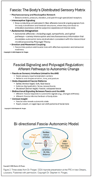
Endings, including mechanoreceptors and nociceptors, have been identified within fascia across the body. These receptors detect mechanical forces such as pressure, stretch, and vibration, as well as signals associated with tissue irritation or injury (see Figure 2).
Interoception refers to the processing of internal bodily signals by the brainstem and higher neural structures. Sensory input originating in fascial tissues contributes to this interoceptive stream through multiple afferent pathways, including spinal and autonomic routes. While the vagus nerve is a major afferent pathway conveying visceral information to brainstem foundational circuits, most somatic sensory input comes from fascia and is transmitted via spinal afferents rather than directly through vagal pathways.
Within this framework, afferent signals from fascia contribute to the ongoing regulation of autonomic state by informing central neural processes involved in evaluating internal and external conditions. Consistent with Polyvagal Theory, this evaluative process – termed neuroception – operates outside of conscious awareness and continuously biases the ANS toward states associated with safety, mobilization, or defensive shutdown.
Integrating these perspectives, fascia can be understood as a distributed sensory interface that contributes to the regulation of autonomic state. Fascial afferents – including mechanoreceptors and nociceptors – detect changes in tension, stretch, and inflammatory processes, and transmit this information through spinal and autonomic pathways to brainstem and subcortical structures. This continuous stream of input contributes to interoception and influences neuroception, the nervous system’s evaluation of safety and threat. In this sense, fascia functions as a source of sensory information that informs central regulatory processes.
Autonomic output, in turn, influences the physiological environment in which fascial tissues operate. Efferent pathways originating in brainstem and spinal cord circuits, and modulated by higher neural structures, regulate vascular tone, immune activity, and metabolic processes that affect tissue properties.
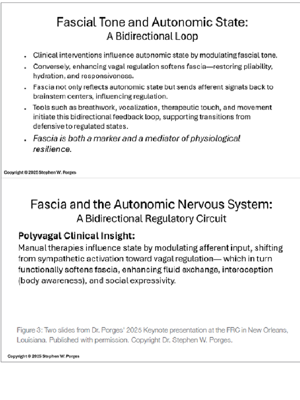
Under conditions of mobilization, sympathetic activity is associated with changes in perfusion and tissue stiffness, while states supporting restoration and social engagement are accompanied by physiological conditions that promote fluid exchange and tissue flexibility.
Thus, rather than a direct bidirectional loop between fascia and the ANS, the relationship is more accurately described as an integrated system in which peripheral sensory input from fascia informs central regulation, and autonomic output shapes the internal milieu of the tissues. Within this framework, fascia is not an autonomous regulator but a dynamic participant in the ongoing coordination of physiological state (see Figure 3).
“Fascia not only reflects autonomic state but sends afferent signals back to brainstem centers, influencing regulation.”
“Tools such as breathwork, vocalization, therapeutic touch, and movement initiate this bidirectional feedback loop, supporting transitions from defensive to regulated states.”
– Porges, 2025 Keynote address at FRC.
Prior to the expansion of Polyvagal Theory through subsequent books and collaborations (Porges 2011, 2017, 2021, 2024; Porges and Porges 2023; Porges and Dana 2018; Porges and Onderko 2025), its foundational concepts were introduced in the 1995 peer-reviewed article, “Orienting in a Defensive World: Mammalian Modifications of our Evolutionary Heritage. A Polyvagal Theory.” In this seminal work, Porges addressed what later became known as the vagal paradox.
Research with human infants revealed that higher levels of cardiac vagal regulation, indexed by respiratory sinus arrhythmia (RSA), were associated with positive health outcomes in full-term newborns. However, in preterm infants, patterns interpreted as vagal-mediated responses were associated with potentially life-threatening events, including bradycardia and apnea. This apparent contradiction raised a fundamental question: how could vagal mechanisms be associated with both adaptive regulation and physiological compromise?
Polyvagal Theory resolves this paradox. As we have already discussed, the vagus is not a unitary system but consists of multiple pathways with distinct anatomical origins and functional properties. For the 1990s, Porges was making a strong challenge to the conventional agonist/antagonist model of ANS function by saying, “the vagus is not one nerve but a family of neural pathways originating in several areas of the brainstem… and at least 80% of the vagal fibers are afferent” (Porges 1995, 304).
The areas of the brainstem of particular relevance are the nucleus ambiguus, which gives rise to the myelinated ventral vagal pathways involved in cardiac regulation and social engagement, and the dorsal motor nucleus of the vagus, which provides unmyelinated efferent pathways associated with more primitive regulatory functions (see Figure 4).
Sensory (afferent) input is processed primarily through the nucleus tractus solitarius. As we already mentioned, these brainstem structures are functionally integrated with other cranial nerve nuclei, including those supporting the social engagement system (cranial nerves V, VII, IX, X, and XI), forming a coordinated network that regulates physiological state and behavior.
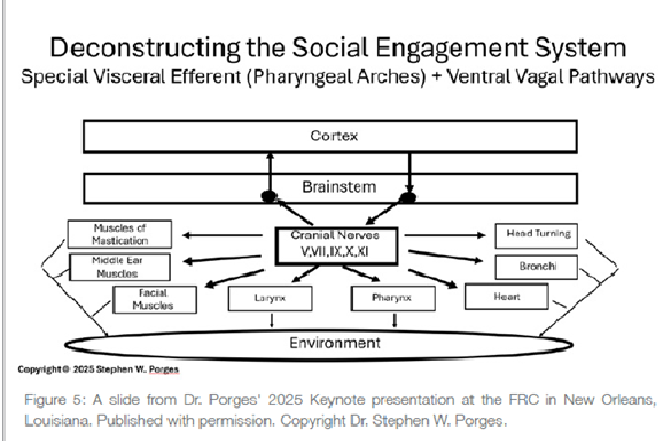
Porges proposed that the ventral and dorsal vagal pathways are functionally distinct, reflecting different evolutionary origins. From this phylogenetic perspective, mammalian brainstem organization includes a ventrally located vagal complex – centered in the nucleus ambiguus – that is integrated with neural circuits supporting communication, emotion, and social bonding. This system enables the coordination of autonomic state with the striated muscles of the face and head, forming the neurophysiological foundation of the social engagement system (see Figure 5).
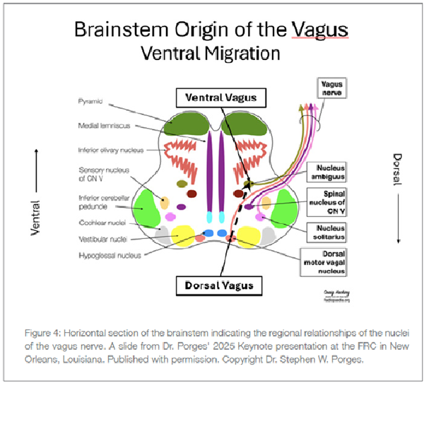
In contrast, earlier-evolving vertebrates rely predominantly on vagal pathways originating in the dorsal motor nucleus, which are associated with more primitive regulatory strategies, including immobilization. While comparisons with reptiles are often used illustratively, it is important to note that modern reptiles are not direct models of ancestral states. Nevertheless, the distinction highlights a key principle: mammalian autonomic regulation includes specialized circuits that support social behavior and the downregulation of defensive states.
As emphasized by Porges, evolution provides an organizing framework for understanding how the ANS supports adaptive behavior. Within this framework, sociality is not merely behavioral but biologically grounded, with cues of safety functioning as powerful regulators of autonomic state. These cues – conveyed through facial expression, vocal prosody, and other features of social interaction – can facilitate physiological states that support health, growth, and restoration.
Polyvagal Theory describes the neurophysiological mechanisms through which social experiences can modulate autonomic state. The ventral vagal pathway, originating in the nucleus ambiguus, is integrated with brainstem circuits that regulate both cardiac function and the striated muscles of the face and head. Through this integration, cues conveyed by facial expression, vocal prosody, and head orientation are linked with autonomic regulation. Signals of safety, detected through these channels, can shift autonomic state toward conditions that support calm engagement, health, and restoration.
The ventral vagal pathway is a core component of the social engagement system, which includes coordinated activity among cranial nerves V, VII, IX, X, and XI. These pathways regulate muscles involved in mastication, facial expression, middle ear function, vocalization, swallowing, and head movement, while also contributing to the regulation of the heart and bronchi. This integrated system enables mammals to both regulate their physiological state and communicate that state to others.
From a developmental perspective, these circuits support early forms of coregulation, as seen in behaviors such as sucking, swallowing, breathing, and vocalization. Through these coordinated processes, the nervous system links autonomic regulation with social interaction, providing a neurophysiological foundation for bonding, communication, and adaptive state regulation.
When the body encounters increased metabolic demands – such as cues of danger, life threat, or illness – autonomic state shifts away from patterns that
Support social engagement. In these conditions, ventral vagal influences on the heart and the striated muscles of the face and head are reduced. This shift may be observed as diminished facial expressivity, reduced vocal prosody, and decreased availability for social interaction. Functionally, this reflects a withdrawal of the ventral vagal “brake” on the heart, allowing heart rate to increase and supporting mobilization in response to challenge.
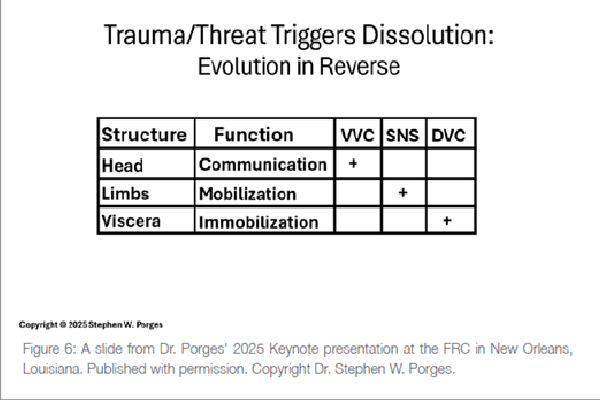
From a polyvagal perspective, mammalian autonomic regulation is organized hierarchically, reflecting phylogenetic development. When conditions are perceived as safe, ventral vagal pathways support social engagement and flexible behavioral responses. If safety is compromised, ventral vagal regulation is reduced, and sympathetic pathways are recruited to support mobilization, including fight-or-flight behaviors. Under conditions of extreme or inescapable threat, more primitive autonomic circuits associated with the dorsal vagal complex may be engaged, contributing to states of immobilization, which can include behavioral shutdown, dissociation, or fainting.
These transitions are not strictly linear or deterministic, but reflect dynamic and context-dependent shifts in autonomic state, mediated by ongoing evaluation of safety and threat through neuroception (see Figure 6).
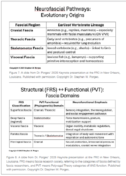
According to Porges, fascia can be considered within an evolutionary framework that parallels the increasing complexity of autonomic regulation, although the relationship is indirect and mediated through neural integration.
In early vertebrates, connective tissues primarily served structural and protective roles, supporting visceral organization and basic physiological regulation. As vertebrates evolved greater motor capacity, connective tissue systems became more differentiated, contributing to the coordination of movement and posture.
With the transition to terrestrial life and the development of pulmonary respiration, additional structural adaptations emerged in the thoracic and diaphragmatic connective tissues, supporting the mechanical demands of breathing.
In mammals, further specialization of cranial and cervical connective tissues is associated with the musculature of the face and head, which is functionally integrated with cranial nerve systems involved in feeding, vocalization, and social communication (see Figure 7).
From a polyvagal perspective, these evolutionary developments can be viewed as increasing the complexity of sensory and motor integration. Rather than viewing fascia as discrete “phylogenetic domains” analogous to vagal pathways, it is more accurate to consider fascia as a distributed system that provides sensory input and mechanical support across multiple functional systems, including those involved in autonomic regulation and social behavior.
Recent fascia research has identified several classifications of fascial tissue, including superficial, deep, visceral, parietal, and meningeal fascia. Porges contrasted these structural categories with the functional organization described in Polyvagal Theory, highlighting potential points of convergence between fascial systems and autonomic regulation (see Figure 8).
Within a polyvagal framework, states associated with safety support spontaneous social engagement and physiological processes related to health, growth, and restoration. These states are mediated by autonomic regulation that influences vascular, metabolic, and immune processes, which in turn affect
The internal milieu of connective tissues, including fascia. Conversely, conditions associated with danger, illness, or life threat are accompanied by shifts in autonomic state that alter these physiological processes, potentially impacting tissue properties such as perfusion, fluid dynamics, and mechanical tension.
Thus, rather than fascia and the autonomic nervous system operating as directly coupled systems, it is more accurate to view fascia as embedded within the physiological environment shaped by autonomic regulation, while also contributing sensory input that informs central processes involved in state regulation.
Fascia and the ANS can be understood as interacting components within an integrated regulatory system that supports adaptive behavior, survival, and social engagement. Rather than forming a direct bidirectional circuit, fascia contributes sensory input through dense networks of mechanoreceptors and interoceptive afferents, while autonomic regulation shapes the physiological environment in which fascial tissues function.
Although parallels can be drawn between fascial organization and the hierarchical framework described in Polyvagal Theory, these relationships are functional rather than structurally mapped. For example, visceral fascia is closely associated with internal organ systems regulated by autonomic processes, skeletomotor fascia is engaged during movement and mobilization, and cranial and cervical connective tissues are involved in structures that support social communication. These associations reflect overlapping functional domains.
______________________________________________________________________________________________________________________
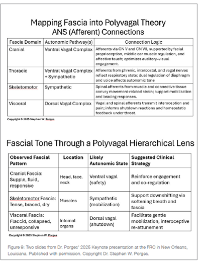
Rather than one-to-one correspondence with specific autonomic pathways. Through its extensive sensory innervation, fascia contributes to the afferent signaling that informs brainstem and subcortical circuits involved in autonomic state regulation (see Figure 9). In this context, fascia can be viewed as a distributed sensory interface participating in the continuous coordination of physiological state.
Porges integrated these perspectives to highlight how physiological state is communicated and regulated within social contexts. From a polyvagal perspective, coregulation emerges through face-to-face interactions in which cues conveyed by facial expression, vocal prosody, and eye gaze are detected by the nervous system as signals of safety. These cues engage the social engagement system and support shifts in autonomic state toward conditions that promote health, growth, and restoration.
Cranial and cervical connective tissues are closely associated with the musculature involved in facial expression and head orientation, which are regulated by cranial nerves, including the trigeminal (CN V) and facial (CN VII) nerves. Through this anatomical proximity, these tissues participate in the expression and transmission of social cues, although they are not themselves components of the neural circuits that regulate autonomic state. When an individual experiences safety, the integrated activity of these neural systems supports expressive vocalization, sustained eye contact, and affiliative behavior.
Respiratory and visceral support structures, including the diaphragm and pelvic floor, play a complementary role in this integrated system. These structures are embedded within fascial networks and are essential for coordinating breathing, intra-abdominal pressure, and visceral function. Through their rhythmic activity and rich sensory innervation, they contribute to interoceptive signaling and influence the physiological conditions under which autonomic regulation occurs. In particular, the coordination of breathing with cardiac regulation – reflected in phenomena such as respiratory sinus arrhythmia – illustrates how these systems are functionally linked at the level of brainstem circuits.
Considering fascia within this framework shifts its role from a purely structural system to a distributed sensory interface contributing to interoceptive and proprioceptive signaling. Rather than directly modulating autonomic tone, fascia provides afferent input that is integrated within brainstem and subcortical circuits involved in neuroception and state regulation. Changes in fascial tension and sensory signaling may therefore reflect, and potentially influence, ongoing shifts in autonomic state.
Fascia can thus be understood as participating in the embodiment of physiological state, contributing to observable patterns in posture, breathing, affect, and behavioral expression. In this way, connective tissue systems and autonomic regulation are functionally interrelated, not as a single bidirectional control circuit, but as components of an integrated system through which physiological state is both regulated and expressed.
What follows is an edited transcript from a conversation recorded on February 19, 2026. Dr. Porges spoke from his home in Florida; Lina Amy Hack from Saskatoon, Saskatchewan. The interview has been condensed and lightly edited for clarity, with Dr. Porges’ approval.
Lina Amy Hack: Good day, Stephen. Thank you for sharing your Fascia Research Congress (FRC) keynote speech notes and slides with us! For readers who may not be familiar with your work, how do you introduce yourself?
Dr. Stephen W. Porges: Hello, and thank you for inviting me to talk with the Rolfers™ and the structural integration community. I’m happy to share my FRC talk and help you edit your summary.
Introducing myself has always been a complex question, because there is no single disciplinary label that accurately describes what I do. My work is truly transdisciplinary, and being a scientist across multiple disciplines doesn’t fit neatly into one category. It requires learning the language and definitions of each field in order to communicate effectively – and that has been part of my journey.
My science is closely tied to that personal journey, which has been an effort to understand what it means to be human – and how that understanding might help us optimize our functioning. In many ways, this parallels the philosophy of Rolfing® Structural Integration: both are concerned with the relationship between structure and function, and between human physiology and behavior. Structure creates constraints, but the challenge is to understand how to function optimally within those constraints.
So, I tend to describe myself as an integrated bio-behavioral neuroscientist – or, more simply, as a curious person.
Lina: The last time we spoke (Hack and Porges 2021), we learned that in the 1980s and 1990s, you helped the then-Rolf Institute® [now the Dr. Ida Rolf Institute®] with research on Rolfing Structural Integration and on how to communicate its clinical relevance to the medical community. Can you orient us to that part of your history for us?
Stephen: That part of my journey with Rolfing [Structural Integration] actually began earlier than the 1980s. It started in the late 1970s when I met with Peter A. Levine, [PhD,] who emerged from the Rolfing community and later developed Somatic Experiencing®. Peter was building a bridge between the impact of trauma on bodily structure and its effects on mental and physical health – helping to integrate those domains as expressions of a common underlying system.
In many ways, Peter was also an early neuroscientist. He was deeply interested in what he referred to as biophysics, particularly the ANS and its responses to stressors and disruptors. That interest led him to seek me out. His perspectives differed significantly from mainstream academic thinking at the time, and he would ask me to help explain the phenomena he was observing clinically. That collaboration marked the beginning of our shared effort to understand processes that we can now describe more formally.
My strength is not as a therapist – I’m not a clinician. But I do have a capacity to deconstruct complex phenomena and identify underlying mechanisms that may be observable. A paper that reflects this perspective is, “Polyvagal Theory: A Journey from Observation to Innovation and Application” (Porges 2025).
I see myself as someone on a journey – respectful of observation, curious about underlying mechanisms, and deeply
Lina: Your keynote — “Fascia and the Autonomic Nervous System: A Polyvagal Framework for Embodied Regulation” – was an extraordinary synthesis. Was it a talk you had been building toward for some time, or did it come together specifically for that audience?
Stephen: I would say it reflects the arc of my scientific journey. The relationship between fascia and the ANS had been percolating in my mind for a long time – I’ve questioned these issues throughout my career. I had been an observer, respectful of what practitioners were doing, and learning from them, but I hadn’t yet fully engaged the underlying mechanistic questions.
When Robert Schleip, [PhD,] invited me to speak at the FRC, I made a decision: it was time to move beyond observation and ask more directly about fascial innervation. To ask, “What is the neural architecture here? And how might that help practitioners understand their clients’ responses?”
I returned to the original literature and approached it from a different perspective – not the primarily correlational autonomic lens I had used previously, but one focused on neuroanatomy: What is the innervation of fascia? And how does it relate to autonomic regulation? What struck me was how much coherence emerged when these findings were interpreted within the ANS framework. It became easier to understand why changes in fascial state are often accompanied by shifts in subjective and physiological experience, and how patterns of bracing or tension may reflect underlying autonomic organization.
My reaction was not only, “This is interesting,” but also, “How did I not fully see this earlier?” I had been engaged with this community for decades. Over time – particularly through listening to individuals describing their experiences of trauma – I had accumulated a broad matrix of observations. The FRC lecture was an opportunity to begin organizing those observations into a more integrated model, linking autonomic regulation with embodied experience, including the role of connective tissue systems.
Lina: You opened your talk by presenting the 1988 research you conducted with Rolfer™ John Cottingham. That was especially meaningful to our community – to see a Rolfer feature at the outset.
Stephen: John Cottingham’s work provided my first close-up view of what Rolfing [Structural Integration] was doing from an autonomic lens. He was deeply interested in understanding the underlying mechanisms. Even at that time, I could see clear autonomic effects emerging from his research (Cottingham, Porges, and Lyon 1988; Cottingham, Porges, and Richmond 1988). Interventions such as the pelvic lift and abdominal massage were associated with measurable shifts in autonomic regulation.
It felt appropriate to begin the talk there. Those early observations provided an important foundation for my later work, and it was also a way of acknowledging and honoring a community that had been exploring these phenomena long before I entered the conversation.
Lina: In the fascia research community, we’ve known for over a decade that fascia is sensory – that it contains mechanoreceptors and nociceptors, and is continuously “listening” to the state of the body. What your talk added for many of us was the other side of the equation. Is fascia also influenced by signals from the brainstem and efferent [motor] pathways of the ANS?
Stephen: Yes. And that’s where the synergy between the sensory properties of fascia and autonomic regulation becomes more apparent. It helps explain why states of safety and calm are often associated with physiological conditions in which tissue may feel more compliant, while states of threat are often accompanied by increased tension.
Fascial sensory information is conveyed through afferent pathways – primarily spinal, with integration in the brainstem and related structures, including regions such as the nucleus tractus solitarius that process visceral input. That’s the afferent [sensory] side of the system. But those signals are part of a larger regulatory process, and what emerges from that integration influences the physiological state of the tissues.
The return pathway is not a dedicated “motor system of fascia” per se. Rather, autonomic efferent pathways regulate vascular tone, immune system activity, and metabolic processes that shape the internal environment of connective tissue. This was a piece that had not been fully integrated into the fascia conversation. There was, at times, an implicit assumption of a self-contained
Communication system within fascia itself. My approach has been guided by parsimony: if a simpler explanation accounts for the observations, we should consider it carefully.
From that perspective, fascia participates in an integrated regulatory process. Sensory input from fascia contributes to central processing, and autonomic output, in turn, influences the conditions under which fascial tissues function. When autonomic state shifts toward mobilization, changes in vascular and neuromuscular activity are often associated with increased tissue stiffness and bracing. When the state supports safety and regulation, those conditions shift toward greater fluidity and flexibility.
Lina: We are talking about fascia as if it were a singular tissue, but in reality, there are multiple types of fasciae.
Stephen: This is where the discussion becomes even more interesting. Consider deep abdominal work directed toward the organs, which engages visceral fascia. Clinically, we observe that visceral tissues can become restricted under conditions of chronic stress or defensive states, much like skeletomuscular systems. In the early work with Cottingham, when deep abdominal techniques were applied, we observed measurable changes in autonomic regulation. These findings suggested that changes in visceral tissue properties are associated with shifts in physiological state.
The fascia research community has already emphasized that muscles cannot be understood independently of fascia. Wherever there is muscle, there is connective tissue that supports and integrates its function, even if anatomical representations often separate them for clarity. Muscle and fascia operate as a functional unit. Extending this perspective, visceral organs are similarly embedded within fascial networks that are responsive to physiological conditions, including those shaped by autonomic regulation.
From a polyvagal perspective, it is useful to consider broad functional domains of motor output, including regulation of visceral organs, skeletomotor activity, and cranial motor systems involved in communication and ingestion. While fascial categories do not map directly onto these domains, there are meaningful functional associations. Visceral fascia is closely related to organ systems regulated
by autonomic processes, skeletomotor fascia participates in movement and postural control, and cranial connective tissues are associated with structures involved in facial expression, vocalization, and head orientation.
Structures such as the diaphragm and pelvic floor are particularly important because they integrate multiple functional demands. They coordinate breathing, support visceral organs, and contribute to postural regulation. These regions include both smooth and striated muscular components and are embedded within complex fascial networks. Because of this, they are especially sensitive to changes in physiological state and are often clinically informative when assessing patterns of tension, breathing, and regulation.
The key point is that fascia participates in an integrated system in which sensory input from connective tissue contributes to central processing, and autonomic regulation shapes the physiological conditions under which these tissues function. Rather than having an independent motor system, fascia reflects and participates in the broader regulatory processes that organize physiological state and behavior.
Lina: And this circles back to the power of a person feeling safe. Are you suggesting that when a person feels safe in the presence of another, it can influence fascial state?
Stephen: Yes. When we consider the relationship between fascia and autonomic regulation through a polyvagal lens, we begin to appreciate how profoundly a sense of safety in the presence of another can influence physiological state. Rolfing [Structural Integration], in many ways, is a pragmatic structural treatment model, and its practitioners are often highly attuned – both to their own internal states and to those of their clients.
Let me take you back to my early work with the Rolfing community. I was asked to develop research methods to evaluate how individuals experienced changes following Rolfing sessions. That led me to begin constructing a set of sensation-based questions that could capture aspects of bodily experience. Over time, this work evolved into what is now known as the Body Perception Questionnaire (BPQ) (see Figure 10).
The questionnaire has since been standardized as a measurement tool, translated into multiple languages, and used with thousands of individuals. It provides a structured way to quantify subjective bodily experience and relate it to underlying physiological processes.
The goal of making the BPQ available to practitioners, including those in structural integration, is to provide a subjective measure of clients’ autonomic reactivity without requiring physiological
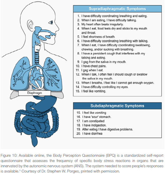
instrumentation. It is a scalable and Accessible tool that can offer meaningful insights into patterns of bodily experience. The Rolfing community is invited to use this resource.¹
Lina: That’s great. I’m thinking about how I might incorporate a questionnaire like this with my clients.
Stephen: The BPQ provides a way to begin assessing aspects of autonomic regulation, even for practitioners who are not formally trained in physiological measurement. It originated from a simple question: “What is happening inside the body during a Rolfing session?” The most direct way to answer that was to ask individuals about their experiences. From those responses, we developed a tool that can be used to better understand patterns of autonomic reactivity.
Using the BPQ, we have also examined how working as a somatic therapist relates to autonomic regulation (Jokić et al. 2023). The measure is sensitive to patterns often associated with trauma history, including indicators of altered autonomic responsiveness. It does not diagnose trauma, but it can reflect how past experiences may be expressed in current physiological regulation.
What we found was that, as a group, body-oriented psychotherapists often report higher levels of adversity experienced early in their lives compared to population averages. Yet, despite this, they frequently demonstrate more effective regulation of their autonomic responses in adulthood. In other words, they are not necessarily more dysregulated; rather, many appear to have developed a greater capacity to modulate their physiological state.
This suggests that individuals drawn to working with trauma may bring both a history of sensitivity and a motivation to support others. Importantly, engaging in this work may also have regulatory benefits. From a polyvagal perspective, therapeutic interactions can function as coregulatory processes – supporting both the client and the practitioner. I find that particularly meaningful, as it highlights how professional engagement can align with both personal history and physiological resilience.
The goal of providing the BPQ to everyone, including practitioners like yourselves, is to give you a subjective measure of your clients’ autonomic reactivity without having to put electrodes on them. It’s a very powerful and scalable, meaning “inexpensive,” tool that is often accessible.
Useful. The Rolfing community is invited to use the links available on my website: www.stephenporges.com/assessments.
Lina: Your study of somatic therapists reflects my experience offering Rolfing sessions for twenty-two years. I have personal practices that help me regulate my nervous system before work, and I carry that into my state while working. I also teach my clients how to do this for themselves. While I’m tired at the end of the day, I also feel genuinely nourished.
Stephen: When you enter the world of somatic therapy, you’re entering a context in which touch is central – but often with individuals who may be highly defensive about being touched. What skilled practitioners bring is not simply technique, but an ability to convey signals of safety through their voice, their proximity, and their regulated presence. These cues enable the client’s nervous system to become more receptive to contact.
From a polyvagal perspective, these signals of safety support shifts in autonomic state that can reduce defensive patterns and allow greater flexibility in physiological regulation. In that context, manual interventions may be experienced as less intrusive and more effective, as the tissues are no longer organized around protective bracing.
What practitioners often describe as a change in tissue quality – greater ease, compliance, or fluidity – can be understood as reflecting changes in the underlying physiological conditions, including vascular dynamics, muscle tone, and autonomic regulation. These are not properties of fascia in isolation, but expressions of an integrated system.
Clinicians frequently report sensing rhythmic changes in tissue during intervention. From a physiological perspective, this may reflect underlying regulatory processes, including cardiovascular and respiratory rhythms, as well as the timing of autonomic responses. These patterns provide indirect information about how the system is adapting and responding over time.
Lina: Studying your FRC slides for this article has given me a new appreciation for what Dr. Rolf herself may have been perceiving when she observed her clients standing and walking. When she said, “Look at the form, look at the structure,” part of what she may have been sensing was the organization of autonomic regulation, even without the language to describe it.
Stephen: I never met Ida, but what I can share is based on conversations with those who knew her and spoke about the way she approached her work. She was an expansive thinker who perceived connections and patterns, including rhythmic processes within the body. The idea of structure as something static – something you simply move through the world with – was likely not the essence of her message.
I think her perspective aligns more closely with what you described in your own experience: becoming present within your body, and through that presence, influencing the experience of others. When practitioners are grounded in this way, they convey cues that can be detected by the nervous system as signals of safety.
In this context, what manual therapists are doing is not simply mechanical. Through movement and changes in tissue organization, they are creating conditions in which clients can experience their bodies outside of defensive states. In a sense, this provides an opportunity for the nervous system to engage in new patterns of regulation – an experiential shift toward a body organized around safety rather than threat.
Lina: When I think about these ideas in relation to Rolfing Structural Integration as a type of training, particularly the Ten Series, it gives me a deeper appreciation for how much Dr. Rolf was perceiving. She seemed to be communicating how to facilitate these changes without the technical language we have now. Was she, in some way, working with autonomic regulation?
Stephen: Yes, and consider how much emphasis she placed on the sacrum, pelvic organization, and breathing. Rolfers are trained to attend closely to the movements of breathing in the chest and throughout the body. For example, when you invite the sternum to move more freely within the thorax, you are indirectly engaging the connective tissues associated with respiration.
The thoracic region is particularly complex because it integrates multiple functional systems, including breathing, posture, and autonomic regulation. When changes occur in this region, practitioners often observe shifts not only in movement but also in facial expression and overall presence. These visible changes may reflect alterations in underlying physiological state, including vascular dynamics and patterns of autonomic regulation.
Clinicians sometimes describe a “brightness” or “glow” in a client’s face following intervention. While the mechanisms are multifactorial, such observations are consistent with shifts toward states that support improved circulation, engagement, and social communication – even when the intervention itself may have involved moments of discomfort. I would expect that practitioners develop their own descriptive language to capture these changes in appearance and expression following treatment.
Lina: Sometimes my clients look like a younger version of themselves – as though years of tension have been lifted and they simply look more like themselves.
Stephen: What you’re observing is that the person’s body appears more open, more accessible – I might even say, more available for connection – when it is no longer organized around threat. They don’t need to be smiling; what matters is that they are accessible.
If we distill compassion to its essence, it is the capacity to be accessible to another. What you are seeing in your clients after a session is a shift in that accessibility – a movement from a state shaped by vulnerability and defense toward one supported by safety. And within that shift, their underlying capacity for connection becomes more visible.
Lina: The vagus nerve has become something of a wellness buzzword. Embedded in both Rolfing and polyvagal language is the assumption that more vagal tone is better. Can you help clarify that?
Stephen: Measures of vagal tone can be informative, but they don’t fully capture how the system is functioning. Vagal activity is dynamic – it increases and decreases as the body responds to changing metabolic demands and supports recovery following those demands.
To address this, I introduced a metric called vagal efficiency. Conceptually, it reflects how effectively vagal pathways regulate the heart in response to challenge. One way to assess this is by observing changes in heart rate across postural shifts – such as moving from supine to seated to standing. These transitions require rapid cardiovascular adjustments, and we can evaluate whether those changes are being mediated efficiently by vagal regulation or whether the system relies more heavily on sympathetic activation.
Across a wide range of clinical conditions, we find that individuals may show reduced vagal efficiency even when traditional measures of vagal tone appear within normal limits or relatively high. In other words, the presence of vagal activity does not necessarily indicate that it is functioning optimally.
This distinction becomes particularly important when considering individuals with trauma histories. Even in cases where experiences do not meet formal diagnostic thresholds, patterns of autonomic regulation may differ. What we observe is that alterations in neural regulation can precede detectable changes in organ function or tissue pathology.
Gastroenterology provides a useful example. Many individuals present with symptoms that are distressing yet do not meet the criteria for structural pathology. In these cases, standard diagnostic approaches may fail to identify a clear cause, leading to a disconnect between the patient’s experience and clinical findings. However, when viewed through the lens of autonomic regulation, these symptoms may reflect disruptions in neural control of organ function rather than damage to the organ itself.
This suggests that dysregulation of the ANS can emerge before structural changes are detectable. Over time, chronic challenges may lead to shifts in regulatory capacity, which can eventually manifest as more persistent physiological or clinical conditions.
Lina: Wonderful insight.
Stephen: You can think of this as a model of numbness – where the body is no longer responding to challenge through the typical pattern of vagal withdrawal followed by mobilization. Instead, the system may no longer be regulating efficiently, and the expected physiological responses are blunted or disorganized.
Within medicine, and more broadly in our culture, there is often an assumption that when something differs from a normative pattern, it must be deficient. A measure increases, decreases, and then increases again – but what does that variation actually represent? That’s really at the core of your question about vagal tone.
As you know, vagal tone is reflected in the patterning of heart rate variability (HRV) – the beat-to-beat changes in heart rate. What matters is not simply the level of vagal activity, but how flexibly and effectively it is regulating physiological state.
Lina: Yes, I explain this to my clients all the time – especially those who have purchased devices that measure “heart rate coherence” but aren’t sure what it means. Let me know if I have this right. This is what I say to them:
“The healthy heart beats irregularly. While it may sound like a regular drum to us, if you really listen to the beat, some beats are far apart and others closer together. It’s subtly speeding up and slowing down all the time – and we want that. That difference between the beats is called ‘variability’, as in, the beat varies.
The sicker we are, the less variability our heartbeat rhythm will have; the healthier we are, the more variability. And, even when we breathe, our heart rate changes. When we breathe in, the rate increases a little bit, and when we exhale, the heart rate decreases a little bit.
This change of rate has been measured for a long time; researchers call it ‘respiratory sinus arrhythmia’, or RSA, also termed ‘heart rate variability’, and clinicians call it ‘heart rate coherence’. And those machines that people buy; they often call it ‘heart coherence’.”
Stephen: That’s a very good summary. And as you mentioned in your introduction, my work in the late 1960s focused on understanding heart rate variability and its neural mechanisms, which led to what later became known as the vagal paradox in newborns. In that research, RSA emerged as a measurable index of vagal influences on the heart, particularly those associated with pathways originating in the nucleus ambiguus.
It’s important to be precise here. RSA is a specific component of heart rate variability that reflects the influence of myelinated vagal pathways on cardiac function. Heart rate variability, more broadly, includes multiple sources of variability, not all of which are vagally mediated.
I do have concerns about how the vagus nerve is often discussed in popular media and even in some professional contexts. The vagus is not a single, uniform pathway.
But a complex system composed of multiple fiber types with distinct functions. It serves as a bidirectional superhighway of neural communication linking the body with brainstem foundational circuits, carrying predominantly afferent signals while also providing efferent pathways that regulate physiological state.
Lina: Let’s get into the ventral vagal and cranial fascia connection. Rolfers are familiar with ventral vagal function as influencing the heart and lungs, and as central to the social engagement system. In your talk, you also linked the ventral vagus to cranial structures.
Stephen: Let me refine part of the framing. The regulation of visceral organs often referred to as “rest and digest” is not exclusively ventral vagal. Much of the ongoing homeostatic regulation of subdiaphragmatic organs is mediated through pathways originating in the dorsal motor nucleus of the vagus. Under typical conditions, these pathways support metabolic and digestive functions. However, under conditions of extreme threat, those same circuits can contribute to energy conservation responses, including reductions in heart rate and, in some cases, syncope.
The ventral vagal pathways, which are unique to mammals, serve a different function. During development, cardioinhibitory neurons originating in more dorsal regions of the brainstem migrate ventrally and become integrated with nuclei that regulate the striated muscles of the face and head. This creates a functional linkage between autonomic regulation and the muscles involved in facial expression, vocalization, and head movement.
As a result, the ventral vagal system coordinates physiological state with social communication. When individuals display prosodic vocal patterns, facial expressivity, and adaptive head orientation, these are observable features of ventral vagal regulation. Conversely, reduced prosody, strained vocalization, or diminished facial expression may reflect a reduction in ventral vagal influence.
The ventral vagal pathways contribute to the regulation of structures such as the larynx and pharynx, while also influencing cardiac function. Through this integrated system, physiological state is both regulated and expressed in ways that support social interaction.
Lina: Okay, to knit this together, the fascia of the face and throat is affiliated with ventral vagal function. We have evolved to reveal our inner comfort or discomfort with our faces?
Dr. Porges: If we put this into evolutionary history, what we realize is that everything about social mammals is built on the social engagement system, which has a specific biological function: it broadcasts the autonomic state of one individual to another. This enables the nervous system of one to assess whether the other is safe to approach, without conscious deliberation. This is the process of neuroception, or detection of threat without awareness.
We experience this in everyday life. You can completely agree with what someone is saying and yet not like them; you feel resistance to them. If you go inside and ask yourself why, what you find is that the person’s intonation, facial expressivity, and gestures weren’t conveying safety or trust. Your nervous system detected that before your cognition did. That is neuroception.
For the Rolfing community, this works in both directions. What your clients’ bodies are broadcasting to you: the rigidity, the lack of prosodic features in their voice, and a high-pitched voice – those are neuroceptive signals of a defensive autonomic state. And what your body is broadcasting to your clients through your presence, your voice, and your quality of attention. When you finish a session, their voice may be very different. You’ll initially attribute it to slower breathing, but then you have to listen a little more carefully and realize that the actual intonation is changing because the ventral vagus also controls the larynx and pharynx. So what you’re getting in the richness of the voice is a higher vagal tone than before.
Lina: Rolfers are working within the skeletomotor system all day long – engaging the fascia of the limbs, trunk, neck, head, hands, and feet. We observe movement patterns, and when clients are in chronic pain, their range of motion is often restricted. What you’ve described aligns with what we see clinically: posture will reflect states of sympathetic dominance – tension, bracing, and reduced tissue compliance. Our interventions aim to reduce these intensities through touch and breath. Are we interacting directly with the sympathetic nervous system?
Stephen: What you are engaging is not the sympathetic system in isolation, but the broader regulatory processes that include ventral vagal influences. The ventral vagal system plays a key role in modulating autonomic state, creating conditions under which defensive activation can be reduced. In many individuals, especially those exposed to chronic stress, ventral vagal regulation may be diminished, resulting in patterns of hypervigilance and persistent mobilization.
From this perspective, your work provides cues that support a shift in state – allowing the nervous system to move away from defensive organization toward greater flexibility and regulation.
Consider the difference between running in fear and running in play. The underlying physiological systems may overlap, but the state is clearly distinguishable through facial expression, vocalization, and posture. These differences are reflected in the coordinated activity of the muscles of the face and head, which provide observable markers of autonomic state.
For practitioners, this becomes part of developing a polyvagal-informed lens: attending to these visible and audible cues as indicators of how the system is organized. While connective tissues are not themselves regulatory structures, they participate in the expression of these states. Patterns of tension, posture, and movement can reflect the underlying organization of the ANS.
When interventions are effective, practitioners often observe increased ease, rhythmicity, and variability in the system. These changes are likely associated with shifts in underlying physiological processes, including autonomic regulation, circulation, and breathing patterns. From a physiological perspective, rhythmicity reflects ongoing regulatory processes – oscillations that indicate the system’s capacity to adapt and return toward homeostasis following challenge.
In this way, manual therapy may facilitate conditions that support the nervous
system’s inherent capacity for regulation, rather than directly imposing change on the system itself.
Lina: What about thoracic fascia?
Stephen: The thoracic region is more complex than purely skeletomotor systems because it integrates multiple functional domains, including respiration, posture, and autonomic regulation. While it can be useful to think in terms of visceral, skeletomotor, and cranial connective tissue systems, these distinctions become less discrete in regions such as the thorax.
As we discussed with structures like the diaphragm and pelvic floor, these areas function as integrative zones – coordinating breathing, visceral support, and movement. Rather than fitting neatly into a single category, they reflect the interaction of multiple systems operating together.
This becomes especially relevant when we consider differences in autonomic state. For example, there is a distinction between immobilization with fear and immobilization without fear. Both may involve stillness, but they reflect very different underlying physiological states. Lying safely in the arms of someone you trust is an example of immobilization that is supported by ventral vagal regulation, whereas fainting or collapse reflects a defensive state associated with dorsal vagal mechanisms.
If movement alone is used as the primary indicator, this distinction can be missed. The observable behavior may appear similar, but the underlying autonomic organization – and therefore the clinical meaning – is fundamentally different.
Lina: And breathing itself – the thoracic fascia is the tissue architecture of breath.
Stephen: Breathing is a portal. This goes back to what you said earlier, that you engage your clients to take a deep breath and exhale slowly. That’s an observation that many people have experienced as useful, and as we have already said, during inhalation, there’s a transitory inhibition of the ventral vagus, and during exhalation, you’re giving permission for the ventral vagus to now resume its inhibitory calming effect.
And that’s why the ratio of inspiration to expiration, when expiration is longer, it’s a more calming experience. When inspiration is greater in that ratio, it’s arousing; it changes your state. Both are functional.
Lina: What about performers – singers, athletes – who are terrified of their audiences but present a convincing ventral vagal face? Are they overriding their nervous system?
Stephen: Those are what I would call superhuman nervous systems – they have developed so much resource that they can sustain the performance, to a point. I’ve worked with a famous opera singer from Europe who lost her voice. Basically, her nervous system gave up; it was no longer going to allow her to sing. Her career changed. She went to graduate school and studied Polyvagal Theory. And in an interesting moment at a conference, I helped her understand what had happened, and she got her voice back.
I also did a podcast with a well-known pop singer who is genuinely petrified by audiences. I asked her how she deals with that. She said, “I turn and look at my band members.” That is co-regulation in action: finding a pocket of safety in an overwhelmingly activating environment.
I have worked with people who survived abduction. One woman was held for nineteen years. She survived not only physically but psychologically intact – a remarkable person with tremendous insight into her own behavior. What allowed her to survive was that she remained an interesting, engaged presence for her captor. Passivity, fawning, and total shutdown – those are not reliable survival strategies. They can provoke escalation rather than diffuse it.
Some people are gifted with a range of nervous system resources that allows them to navigate extreme situations. And that word gifted matters: it is not a choice, not an intention. If the resource isn’t there, the body shuts down. An appeaser has much greater range until the body says, “I’ve had enough, I’m out of here.”
Lina: Rolfers often receive clients who have seen every other practitioner, and they come into our offices saying, “What else could you possibly do?” And the Rolfing Ten Series® does help.
Stephen: They’re in the door – that’s the important thing. They’ve arrived, and that reflects a continued search for change.
Let me offer a way to frame this. Many of these individuals have experienced persistent patterns of physiological regulation shaped by stress or trauma. These patterns can be expressed in the body through tension, restricted movement, and altered autonomic state.
Connective tissues participate in these patterns, reflecting how the system has adapted over time.
Signals of safety – conveyed through your presence, attunement, and interaction – can support shifts in autonomic state. For some individuals, that may be sufficient to begin changing how their body is organized. For others, additional forms of intervention may be helpful.
This is where manual therapy plays an important role. Through skilled touch and guided interaction, practitioners can create conditions in which clients experience changes in tension, movement, and internal state. Even if these changes are initially transient, they provide an important reference point – an experience of the body organized differently.
Once a client has experienced that shift, it becomes meaningful. It is often recognized as familiar at some level, even if it has not been previously accessible. With repeated experiences, individuals may develop great capacity to access and sustain these states.
Over time, this process can support meaningful changes in how a person experiences their body – greater ease, increased accessibility, and a reduced tendency to organize around defensive states.
Lina: Manual therapists are giving the fascia a new message from the ANS through social interaction as somatic therapists and through contact with our hands.
Stephen: Remember the important message I have for the fascia community: fascia is a tissue system whose properties are shaped by autonomic regulation.
Practices that support a sense of safety may influence autonomic state, which in turn can affect the physiological conditions under which fascial tissues function. So, in a way, you could have hands-off Rolfing [Structural Integration] and hands-on Rolfing [Structural Integration], in both, you’re shifting the fascia through a relationship with your client.
Even though there’s great relief after one session, because of the way that individual interacts with the world, the pain pattern is reinforced in one way or another. Then their challenge likely can’t be ‘fixed’ with only the singular visit, and the person comes back. The toxicity of their life has not been adjusted. So there’s learning that takes place. Yes we can help you experience this new, easier state, but there’s a part of feeling better that you have to do with your own nervous system.
I use the word navigate, we each have to learn to navigate and respect our world, our environment.
We have this very gifted nervous system that can deal with threats miraculously, as long as the threat is not twenty-four hours a day, seven days a week. Our nervous system cannot deal with threats all the time. For some people, they just need a good night’s sleep, or time with a loved one, or a pet that enables their body to recover. Other people need more. When we respect and honor our body’s needs, this is what we realize. It’s not wrong to be under stress, and you can do very well with it. But you can’t do it all the time. You need to have periods of time of what I like to call – social nourishment.
A Working Definition
Lina: Your FRC talk subtitle was “A Polyvagal Framework for Embodied Regulation.” What does embodied regulation mean to you?
Stephen: Over the course of my work trying to understand how the body responds to trauma, I’ve come to see that recovery is, in many ways, a process of re-embodiment. It is a return to being able to experience the body as a place that feels accessible, organized, and safe.
When individuals have been living in states of chronic threat, their experience of their body can become altered – constrained, defensive, or even disconnected. From a polyvagal perspective, embodiment reflects the capacity to remain present within one’s body while the ANS supports regulation rather than defence.
Practitioners who work with fascia are often engaging with tissues that reflect these patterns of adaptation – tension, restriction, and altered movement.
Through their work, they can help create conditions in which clients begin to experience their bodies differently. That shift can be understood as an initial step toward re-embodiment.
But that is only the beginning. The next question becomes: what supports the individual in sustaining that state? What forms of nourishment – physiological, relational, and environmental – allow the nervous system to remain in a state of accessibility for longer periods of time?
Lina: That makes so much sense; their environment is setting the tone. Yet we can plant a seed, so to speak. One of your slides read, “Fascia enables the nervous system to translate internal physiological signals into embodied behavior, shaping posture, breath, affect, and presence.” When we invest in wellness that allows for a peaceful and curious nervous system, how we move, feel, and communicate will have this tone. And that’s how we help ourselves, and the people around us. Your research has this positive message from years and years of work – thank you!
Stephen: This perspective didn’t emerge in isolation. It developed through a long and collaborative journey – beginning with Peter Levine, continuing through my work with John Cottingham, and more recently through conversations with Robert Schleip, as well as contributions from Jim Oschman [author James L. Oschman, PhD], Stanley Rosenberg [practitioner and author], and Pat Ogden [PhD]. All of them shared a commitment to understanding the phenomena observed in the Rolfing paradigm and other somatic therapies.
Peter was asking these questions as early as the 1970s. He wanted to understand why individuals exhibited particular physiological and behavioral responses following trauma. That was a reasonable and important question, and it initiated a line of inquiry that has continued to evolve.
What has changed over time is that the language and the science have begun to converge. Concepts such as “embodied regulation,” which may once have been difficult to articulate within scientific contexts, are now more widely accepted. We are increasingly able to describe these observations in terms that are meaningful both to clinicians and to researchers.
With respect to fascia, it is not that the tissue itself carries complex “messages” in isolation. Rather, it participates in a broader system in which neural pathways, vascular dynamics, and mechanical properties interact. As our understanding of these interactions has grown, the discussion has become more precise and more integrative.
Lina: It paints a deeper picture about what “opening” tissue is doing – that it’s not just relaxation, but a shift in physiology.
Stephen: Yes. If we think of fascia as a richly innervated tissue contributing to sensory input, then manual therapy can be understood as influencing the signals being conveyed to the nervous system. Rather than viewing fascia as simply “releasing,” we can consider how changes in tissue tension and movement alter the afferent information reaching brainstem regulatory circuits.
From that perspective, shifts in tissue organization are accompanied by changes in how the body is being monitored and regulated. These changes can, in turn, influence autonomic output, including processes such as circulation and visceral regulation.
I experienced this personally when I went through the Rolfing Ten Series®. I remember noticing changes in my own face following sessions – a kind of brightness or “glow.” At the time, I began to think about what might underlie that observation. It likely reflects changes in physiological state, including vascular dynamics and autonomic regulation, rather than a purely local tissue effect.
When practitioners observe their clients, they are often sensing patterns within a highly interconnected system. Fascia can be thought of as part of a broader sensory interface contributing to interoception, providing continuous input that informs autonomic regulation. Through this lens, it participates in the processes that support health, adaptation, and social engagement.
One of the contributions of Polyvagal Theory is to emphasize that sociality itself is a biological function. When physiological state supports safety, individuals spontaneously engage with others, and coregulation emerges. These processes are not primarily driven by cognition, but by underlying neural regulation – they are experienced, rather than consciously constructed.
Lina: Your work and your talk at the FRC feel like the closest thing to a “theory of everything” to explain Rolfing Structural Integration I’ve encountered.
Stephen: Thank you. What you’re observing is really a reflection of my journey. I’m a curious person, and understanding the human experience.
has always been my central focus. Over the decades, I’ve been refining that understanding – trying to identify the underlying principles that support how we function, adapt, and connect.
In that sense, it becomes less about any single discipline and more about understanding how to optimize the human experience. When we begin to think in those terms, it naturally extends into many domains: how we structure schools, how we design hospitals, how we organize work environments, even how we think about our built spaces.
At its core, this is an effort to understand what the human body requires to feel safe, to engage, and to thrive – and what it does not tolerate well. Living in our world comes with a cost when the nervous system is asked to remain in a state of defense for too long. The challenge is not avoiding that cost entirely, but learning how to restore balance through experiences of safety, connection, and social nourishment.
Lina: Social nourishment; that’s brilliant. That is going right into my pocket. That brings a natural closure point for our conversation, so much to ponder. I’ve enjoyed our conversation very much.
Stephen: As you can tell, I’m not a passive participant in interviews; I’m engaged and enjoying it. Curiosity is extremely important in life, and the passion to learn and understand. It’s not just the accumulation of knowledge; it’s how that knowledge can help you understand the human experience.
Lina: Yes – and to apply that knowledge to the one individual client in front of us. That’s where the art meets the science.
Stephen: Yes, it is an art. We are part of a community of practitioners who have chosen to engage in this work – individuals who are both skilled and sensitive, working with some of the most vulnerable aspects of human experience. And they are doing so with care and effectiveness. There is much we can learn from that.
Lina: Thank you for helping us learn from you! Take care.
Stephen: You are welcome, nice job.
Stephen W. Porges, PhD, is a behavioral neuroscientist whose research has transformed understanding of how the autonomic nervous system shapes social behavior, emotion, and health. He is the originator of Polyvagal Theory, first introduced in 1995, a framework describing how brainstem autonomic circuits regulate responses to safety and threat and support social engagement through the ventral vagal complex – an evolutionarily derived neural system linking autonomic regulation with the neural control of the face, head, and voice.
Dr. Porges is a Distinguished University Scientist at the Kinsey Institute, Indiana University, where he is the founding director of the Traumatic Stress Research Consortium. He also serves as Professor of Psychiatry at the University of North Carolina at Chapel Hill and Courtesy Professor in the Department of Psychiatry at the University of Florida College of Medicine in Jacksonville. He holds emeritus professorial appointments at the University of Maryland and the University of Illinois College of Medicine.
He has authored more than 400 peer-reviewed publications, cited in over 65,000 scientific works. He has written several books translating autonomic science for clinical and public audiences, including Our Polyvagal World (Porges and Porges 2023) and Safe and Sound (Porges and Onderko 2025).
His translational work includes the Safe and Sound Protocol™, a structured listening intervention he developed, and Sonocea® Sonic Augmentation Technology™ (SAT™), an acoustic neuromodulation platform codeveloped with Anthony Gorry. The Rest and Restore Protocol™ is an application of this platform designed to support autonomic regulation and restorative states. His work has also led to patented methods for monitoring and modulating autonomic function.
Lina Amy Hack, BS, BA, SEP, became a Rolfer™ in 2004 and is now a Certified Advanced Rolfer™ (2016) practicing in Canada. She has an honors biochemistry degree from Simon Fraser University (2000) and a high-honors psychology degree from the University of Saskatchewan (2013), as well as a Somatic Experiencing® Practitioner (2015) certification. Hack is the Editor-in-Chief of Structure, Function, Integration.
Bear, Matthew, H., Vamsi Reddy, and Pradeep C. Bollu. 2022. Neuroanatomy, hypothalamus. Available from https://www.ncbi.nlm.nih.gov/books/NBK525993/.
Cottingham, J. T., Stephen W. Porges, and T. Lyon. 1988. Effects of soft tissue mobilization (Rolfing pelvic lift) on parasympathetic tone in two age groups. Physical Therapy 68(3):352-356.
Cottingham, J. T., Stephen W. Porges, and K. Richmond. 1988. Shifts in pelvic inclination angle and parasympathetic tone produced by Rolfing soft tissue manipulation. Physical Therapy 68(9):1364-1370.
Jokić Biljana, Danka Purić, Herbert Grassmann, Christopher G. Walling, Evan J. Nix, Stephen W. Porges, and Jacek Kolacz. 2023. Association of childhood maltreatment with adult body awareness and autonomic reactivity: The moderating effect of practicing body psychotherapy. Psychotherapy 60(2):159-170.
Hack, Lina Amy, and Stephen W. Porges. 2021. Polyvagal theory and trauma: An interview with Stephen Porges, PhD. Structure, Function, Integration 49(2):18-25.
Lechan, Ronald M. and Roberto Toni. 2016. Functional anatomy of the hypothalamus and pituitary. Available from https://www.ncbi.nlm.nih.gov/sites/books/NBK279126/.
Porges, Stephen W. 2025. Polyvagal theory: A journey from physiological observation to neural innervation and clinical insight. Frontiers in Behavioral Neuroscience 19:1659083.
___. 2024. Polyvagal perspectives: Interventions, practices, and strategies. New York: Norton Professional Books.
___. 2023. The vagal paradox: A polyvagal solution. Comprehensive Psychoneuroendocrinology 16:100200.
___. 2021. Polyvagal safety: Attachment, communication, self-regulation. New York: Norton Professional Books.
___. 2017. The pocket guide to the polyvagal theory: The transformative power of feeling safe. New York: Norton Professional Books.
___. 2011. The polyvagal theory: Neurophysiological foundations of emotions, attachment, communication, and self-regulation. New York: Norton Professional Books.
___. 2007. The polyvagal perspective. Biological Psychology 74(2):116-143.
___. 1995. Orienting in a defensive world: Mammalian modifications of our evolutionary heritage. A polyvagal theory. Psychophysiology 32(4):301-318.
Porges, Stephen W. and Deborah A. Dana (Eds). 2018. Clinical applications of the polyvagal theory: The emergence of polyvagal-informed therapies. New York: Norton Professional Books.
Porges, Stephen W. and Karen Onderko. 2025. Safe and sound: A polyvagal approach for connection, change, and healing. Boulder, Colorado: Sounds True.
Porges, Stephen W. and Seth Porges. 2023. Our polyvagal world: How safety and trauma change us. New York: Norton Professional Books.
Suarez-Rodriguez, Vidina, Caterina Fede, Carmelo Pirri, Lucia Petrelli, Juan Francisco Loro-Ferrer, David Rodriguez-Ruiz, Raffaele De Caro, and Carla Stecco. 2022. Fascia innervation: A systematic review of the literature. International Journal of Molecular Sciences 23:5674.
Waxenbaum, Joshua A., Vamsi Reddy, and Joe M. Das. 2025. Anatomy, autonomic nervous system. Available from https://www.ncbi.nlm.nih.gov/sites/books/NBK539845/.
Polyvagal Theory; autonomic nervous system (ANS); fascia; vagus nerve; ventral vagal; dorsal vagal; neuroception; interoception; social engagement system; coregulation; fascia as sensory organ; mechanoreceptors; nociceptors; cranial fascia; visceral fascia; skeletomotor fascia; heart rate variability, respiratory sinus arrhythmia; vagal tone; vagal efficiency; sympathetic nervous system; parasympathetic nervous system; face-heart connection; embodied regulation; trauma imprints; body perception questionnaire (BPQ); Rolfing Structural Integration; brainstem; phylogenetic evolution; diaphragm; pelvic floor; safety. ■